Much of histioteuthid systematics is based on photophore patterns. Details of these patterns, however, were not carefully examined previously which has contributed to some confusion in identification. Patterns seen here are for species with 3 or 4 series of photophores on the arms and with photophores of more-or-less uniform size (11 species). Species with more photophores series on the arms (4 species) have much more variable patterns and the differences between individual and interspecific (or higher) variation has yet to be unraveled. Species with photophores of mixed sizes (3 species) pose problems of which photophores to include in the patterns; these problem remain to be solved.
Terminology:
The drawing below shows the basic photophore pattern for compound photophores of histioteuthid squids. The drawing represents a head rolled out on either side, like the unrolling of a cylinder, with the ventral surface of the head in the center and the dorsal midline on either side of the drawing. The light brown circles represent the photogenic region of each photophore with its surrounding reflectors and pigment but not the distal reflecting chamber or color filter. The photophores have been joined by lines in order to identify various photophore groups.

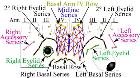
Figure. Histioteuthid head photophore pattern showing terminology. Drawing by R. Young.
The matrix of red lines on the ventral surface of the head (the Ventral Matrix) is defined by (1) the Basal Arm IV Rows (yellow lines) which are the most posterior rows on arms IV that clearly forms a straight line of the orientation shown - photophores in each of these rows also define the number of photophore series on each arm, (2) the Right and Left 2° Eyelid Series (black line) and (3) the Midline series (thick blue line). The matrix is most easily recognized by starting with the Midline Series. Next connect photophores by red lines that form diamond-shapes. Blue lines, then, connect the anterior and posterior poins of each diamond. The resulting matrix is bilaterally symmetrical except for a single "rogue" photophore colored red. Individual photophores within these series are identified by numbering from medial to lateral or, if not paired (e.g., the Basal Row, from viewers left to right.
Construction of the Ventral Matrix uses a few simple rules: The Basal Row and the Right and Left Basal Series are not part of the matrix. The matrix is formed by oblique (red) lines connecting photophores in diamond patterns. Longitudinal (blue) lines connect anterior and posterior photophores within a red diamond. Lines (red and/or blue) never cross one another except at a node (ie, a photophore). The oblique (red) lines can only connect to a photophore of a 2° eyelid series if a longitudinal (blue) line can connect to the same photophore.
Head photophores: Type 1
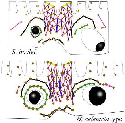
Figure. Stylized drawing of the head photophore patterns. Top - Stigmatoteuthis hoylei, based on specimens from Hawaiian waters. Bottom - Histioteuthis celetaria-type, based on specimens of H. sp. A from Hawaiian waters. Drawings by R. Young.
- The Basal Arm IV Row has 3 photophores.
- The Midline Series has 3 photophores.
- The Ventral Matrix has 9 longitudinal (blue) series and 38 photophores.
- Photophores of the Ventral Matrix not aligned in transverse rows.
- The Right Eyelid Series has 17 photophores.
- The Left Eyelid Series has 6 photophores.
- The 2° Right Eyelid Series has 8 photophores.
- The 2° Left Eyelid Series has 7 photophores.
- The Right Accessory Series has 2 photophoes.
- The Left Accessory Series has 3 photophores.
- All of the Type 1a features listed above.
- An additional posterior photophore, a "rogue" photophore colored red, lies ("dangles") just outside the matrix (it was left out for emphasis), but is connected by a blue line.
Head photophores: Type 2
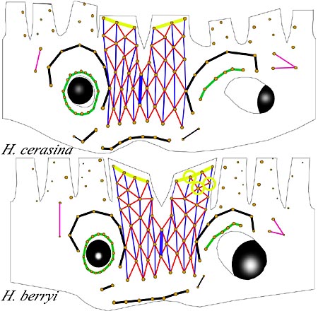
Figure. Stylized drawing of the head photophore patterns. Top - Histioteuthis cerasina, based on specimens from Hawaiian waters. Bottom - Histioteuthis berryi, based on specimens from Hawaiian waters. We thank M. Seki, NMFS, for providing a specimen that was in good condition that enabled confirmation of the photophore pattern. Drawings by R. Young.
- The Basal Arm IV Row has 3 photophores.
- The Midline Series has two photophores.
- The Ventral Matrix has 11 longitudinal (blue) series and 42 photophores.
- Photophores of the Ventral Matrix aligned in transverse rows.
- The Right Eyelid Series has 17 photophores in both.
- The Left Eyelid Series has 6 photophores in both.
- The 2° Right Eyelid Series has 8 photophores in both.
- The 2° Left Eyelid Series has 7 photophores in both.
- The Right Accessory Series has 2 large photophores in both.
- The Left Accessory Series has 3 photophores in both.
Note that the bottom 5 characteristics are the same as in Type 1. That is, the first four characteristics differentiate the types.
The Type 2a pattern, found only in H. berryi (drawing on the right), has the following features:
- All of the Type 2a features listed above except that:
- The Basal Arm IV Row has 4 photophores.
- The Ventral Matrix has 3 additional photophores on either side (circled in yellow on one side) giving the Ventral Matrix 48 photophores. Otherwise, the Ventral Matrix of Types 2a and 2b are the same.
Defining the photophore patterns, as done here, allows more accurate descriptions and fewer errors in identification. We are impressed by the great similarity in number of photophores in the different photophore groups between species. The Basal photophores seem especially important in species identification. We suspect that new characters will be found in the relative positions of some photophores within groups. A good example of this is the sawtooth pattern of the Basal Row with three teeth, as seen in Stigmatoteuthis hoylei, which contrasts with the single sawtooth in Histioteuthis sp. A and the absence of sawteeth in H. pacifica.
Mantle photophores:
Here we point out the extreme consistency the the number of photophores near the anterior mantle margin. We have selected three species with different mantle shapes and photophore sizes. Histioteuthis sp. A has a cyclindrical mantle with large photophores. Stigmatoteuthis hoylei has a conical mantle with photophores of intermediate size and Histioteuthis cerasina an intermediate mantle shape with small photophores.
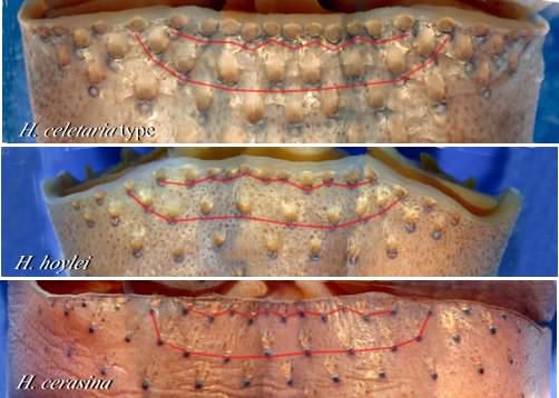
Figure. Composite photographs showing the arrangement of anterior mantle photophores in three species, Histioteuthis celetaria-type (H. sp. A), 25 mm ML, Hawaiian waters, Stigmatoteuthis hoylei, 25 mm ML, Hawaiian waters, Histioteuthis cerasina, 60 mm ML, Hawaiian waters. Photographs by R. Young.
The photographs above are montages each taken from a series of different angles to show nearly the entire anterior mantle margin (ie, ventral, lateral and dorsolateral). We have highlighted the first two photophore rows with a light red line. All three species have 11 photophores in the first series (Ventral Marginal Row) and 10 in the second (2° Marginal Row). The Ventral Marginal Row runs between the outer margins of the mantle locking-apparatuses. However, it is usually easiest to locate the starting and ending points by first noting where the 2° Marginal Row abuts the anterior mantle margin.
The third row of photophores (not hightlighted) appears to have 11 photophores in each species but the identification of photophores as belonging to a row is becoming more difficult.
The total number of photophores on the mantle varies greatly among species. A reliable, objective method for determining the number of rows, etc. of these photophores has yet to be found.


