


Histioteuthis macrohista
Richard E. Young and Michael Vecchione

Introduction
H. macrohista, along with H. bonnellii, are the only members of the genus with a very large inner web. H. macrohista is apparently much the smaller of the two with all known individuals less than 70 mm ML while H. bonnellii is known from individuals as large as about 330 mm ML. The function of the deep web is unknown. However, its heavy pigmentation would make it an effective tool in concealing the bioluminescence emitted by struggling prey. A prudent predator, in this dark habitat, does not want to attract the attention of larger animals. If true, one wonders why all histioteuthis haven't adopted this strategy.
Characteristics
- Photophores
- Compound photophores number 16 (rarely 15) around right eye.
- 2 large, round, broadly-separated, dark photophores on left posterior margin of ventral surface of head (see drawing to the right).
Click on an image to view larger version & data in a new window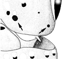
Figure. Left posteroventral corner of head of H. macrohista, 19 mm ML,46°S 147°E, showing the two diagnostic dark photophores. Drawing from Voss, et al. (1998).
- Buccal crown
- Buccal crown with 7 buccal supports.
Comments
More details of the description can be found here.Species of the bonnellii-group are distinguished by the following characteristics:
- Photophores
- Single, elongate, simple photophore at end of each arm I-III.
- Type 1b head photophore pattern (needs confirmation from H. macrohista).
- Two or three large, round photophores on left posteroventral margin of head.
- Compound photophores of large, uniform size on anterior half of ventral mantle.
- Arms IV with 3 longitudinal series on arm base and without separated group of compound photophores at arm tips.
- Web
- Deep inner web between arms I-III (>50% of length of longest arm; no other species have webs > 30%).
- Buccal membrane attachments
- Multiple attachments of the fourth (ventral) supports of the buccal crown (i.e., 1 each to sides of arms IV and to junctures of web segments from arms III and IV).
- Tubercles
- Absent
This species is most easily separated from its close relative, H. bonnellii by the presence of 7, rather than 6, supports in the buccal crown, 16 rather than 17 photophores on the right eyelid (numbers rarely overlap) and the presence of 2 rather than 3 conspicuous, dark, round photophores on the posteroventral margin of the head on the left side. An additional difference is the manner that the inner webs between the third arms join those of the ventral arms. In H. macrohista the two junctions are widely separated, while in H. bonnellii they nearly form a common junction. Voss, et al. (1998) also show differences in sperm mass length and number of loops in the ejaculatory apparatus of the spermatophores, in the number of gill lamellae as well as the presence or absence of an elongate, simple but relatively small photophore on the tips of arms IV.
The above description, comparisons and comments, with the exception of the head photophore pattern, is taken from Voss, et al., 1998.
We list the head photophore pattern for the bonnellii-group as Type 1b. We have only examined H. bonnellii, however, drawings in Voss (1969, the drawing used here in the title illustration) suggest that H. macrohista also has this pattern.
Life History
Females mature at 49->65 mm ML; males mature at 40-55 mm ML.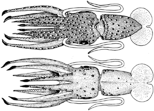
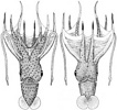
Figure. Dorsal and ventral views of a young H. macrohista, 11 mm ML, 40°S 161° E. Note the presence of the arm-tip photophores at this small size (this can be easily seen squid as small as 6 mm ML) but the low depth of the inner web. Drawing from Voss, 1969 (Figs. 37b,c).
Distribution
Vertical distribution
Little reliable information is available on the vertical distribution of this species; however, presumably, it inhabits mesopelagic waters at least as a subadult.
Geographical distribution
Type locality: Tasman Sea, 45°10'S, 160°10'E. H. macrohista primarily occupies the region near the Southern Subtropical Convergence. Voss et al., (1998) suggest that the absence from the eastern Pacific is real and not a sampling artifact. In the well-sampled Atlantic range of this species, adult males were taken close to continental slopes and in the open ocean.
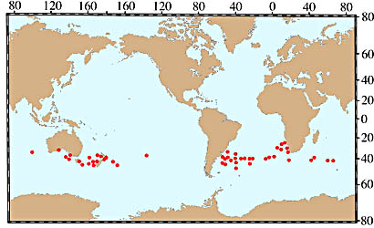
Figure. Chart of the geographical distribution of H. macrohista. Chart modified from Voss, et al. (1998).
References
Voss, N. A. 1969. A monograph of the Cephalopoda of the North Atlantic: The family Histioteuthidae. Bull. Mar. Sci., 19: 713-867.
Voss, N.A., K. N. Nesis, P. G. Rodhouse. 1998. The cephalopod family Histioteuthidae (Oegopsida): Systematics, biology, and biogeography. Smithson. Contr. Zool., 586(2): 293-372.
Title Illustrations
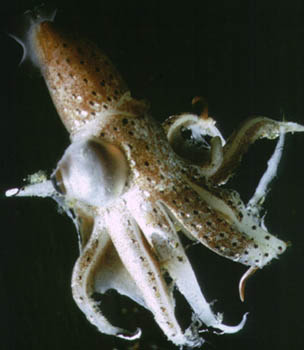
- Ventral view of a damaged squid; note arm-tip photophores and deep web. Photograph by E. McSweeny.
- Ventral and dorsal views of holotype, 52 mm ML, female, 45° 10'S, 160° 10'E. Drawing from Voss, 1969 (Figs. 36a, 37a).
Other illustrations
- entral and dorsal views of a young squid, 40°S 161° E. Drawing from Voss, 1969 (Figs. 37b,c).
- Distribution chart: Modified from Voss, et al. (1998).

| Copyright | © 2000 E. McSweeny |
|---|
| Copyright | © 1969 Bulletin of Marine Science |
|---|
About This Page

University of Hawaii, Honolulu, HI, USA
National Museum of Natural History, Washington, D. C. , USA
Page copyright © 2013 and
 Page: Tree of Life
Histioteuthis macrohista .
Authored by
Richard E. Young and Michael Vecchione.
The TEXT of this page is licensed under the
Creative Commons Attribution-NonCommercial License - Version 3.0. Note that images and other media
featured on this page are each governed by their own license, and they may or may not be available
for reuse. Click on an image or a media link to access the media data window, which provides the
relevant licensing information. For the general terms and conditions of ToL material reuse and
redistribution, please see the Tree of Life Copyright
Policies.
Page: Tree of Life
Histioteuthis macrohista .
Authored by
Richard E. Young and Michael Vecchione.
The TEXT of this page is licensed under the
Creative Commons Attribution-NonCommercial License - Version 3.0. Note that images and other media
featured on this page are each governed by their own license, and they may or may not be available
for reuse. Click on an image or a media link to access the media data window, which provides the
relevant licensing information. For the general terms and conditions of ToL material reuse and
redistribution, please see the Tree of Life Copyright
Policies.
Citing this page:
Young, Richard E. and Michael Vecchione. 2000. Histioteuthis macrohista . Version 01 January 2000 (under construction). http://tolweb.org/Histioteuthis_macrohista/19787/2000.01.01 in The Tree of Life Web Project, http://tolweb.org/


