- Photophores (head)
- Compound photophores (Type 1b):
 Click on an image to view larger version & data in a new window
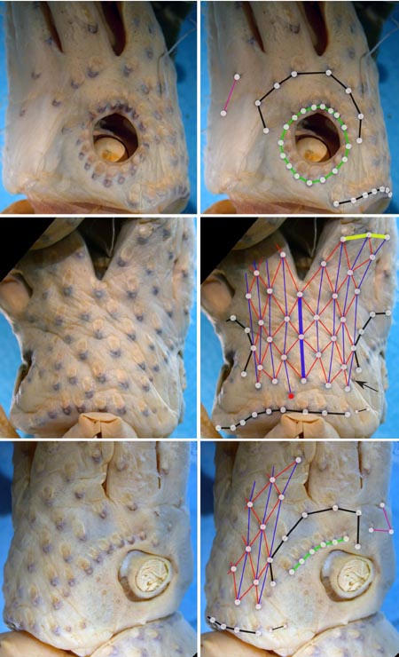
Figure. Various views of the head photophores of H. pacifica, holotype, 74 mm ML, female, preserved. Right photographs - Show one or more of the basic photophore groups, from different views, with superimposed lines and dots to emphasize the photophore groups. Left photographs - Show the same pictures without superimposed lines and dots so that the photophores can be seen without obstruction. The superimposed lines are formed by first placing white dots over the pigmented, photogenic cups of all photophore in a particular group. The dots are then joined by a line of the appropriate color. Photographs by R. Young.
- Basal Arm IV Row with 3 photophores. Medial photophore separated by 1 photophore from anterior photophore of Midline Series.
- Midline Series with 3 photophores.
- Right Eyelid Series with 17 photophores .
- Left Eyelid Series with 6 photophores.
- 2° Right Eyelid Series with 8 photophores.
- 2° Left Eyelid Series with 7 photophores.
- Ventral Matrix with 36 photophores including an asymmetrically placed dangling, rogue photophore (colored red in illustration), and 9 longitudinal (blue) series. Attachment to 2° Eyelid Series at 2° eyelid photophores no. 2 either side.
- Ventral Matrix photophores do not align in transverse rows.
- Right basal series absent.
- Left basal series apparently with two small photophores.
- Basal row with 9 photophores smoothly aligned (i.e. without a sawtooth).
- Right Accessory Series with 2 photophores (see below).
- Left Accessory Series with 3 photophores (see below).
Click on an image to view larger version & data in a new window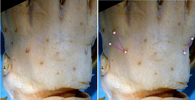
Figure. Dorsal view of the head photophores of H. pacifica, holotype. Photographs by R. Young.
- Simple photophores (round, black dots):
- Arrangement as illustrated.
- Large, simple photophores on dorsal surface of head absent.
- Small, simple photophores not easy to recognize and may be variable.
- Compound photophores (Type 1b):
- Photophores (arms IV)
- Arms IV with 3 series of compound photophores in proximal half.
- Group of small, compound photophores at arm tip, separated by a gap from other arm photophores.
Click on an image to view larger version & data in a new window
Figure. Ventral view of left arm IV of H. pacifica. Drawing extracted from Fig. 17a of Voss, 1969.
- Photophores (mantle)
- Compound photophores of large, uniform size and evenly spaced on nearly all of ventral mantle.
Click on an image to view larger version & data in a new window
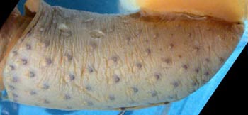
Figure. Mantle photophores of H. pacifica. Top - Ventral view of mantle, 74 mm ML. Drawing a portion of Fig. 17A from Voss, 1969. Bottom - Ventrolateral view of mantle, holotype. Photograph of preserved squid by R. Young.
- Tentacles
- Manus with suckers in 6-7 irregular series. Drawing and photograph from holotype.
Click on an image to view larger version & data in a new window

Figure. Oral view of the tentacular club, holotype, 74 mm ML. Top - Drawing from Voss, 1969 (Fig. 17g). Bottom - Partial left club, preserved. Image inverted making it appear as the right club in order to match the orientation of the drawing. Photograph by R. Young.
- Arms
- Length 80-130% of ML; arms II and III longest.
- Large arm suckers smooth.
- Occipital folds
- Two prominent occipital folds present on each side of head.
- Two prominent occipital folds present on each side of head.
- Web and buccal crown
- Inner web vestigial; outer web absent
- Buccal crown with 7 supports.
Click on an image to view larger version & data in a new window
Figure. Oral view of buccal crown of H. pacifica, 74 mm ML. Drawing from Voss, 1969 (Fig. 17f).
- Funnel organ
- Dorsal pad of funnel organ unsculptured
Click on an image to view larger version & data in a new window
Figure. Ventral view of the funnel organ of H. pacifica, 74 mm ML. Drawing from Voss, 1969 (Fig. 17e).
- Fins
- Length 43-60% of ML; width 63-75% of ML.
- Length 43-60% of ML; width 63-75% of ML.
- Gladius
- Anterior shoulders of vanes broadly rounded.
Click on an image to view larger version & data in a new window
Figure. Ventral view of gladius of H. pacifica, paratype, mature male, 51 mm ML, 13° 49'N, 120° 42'E. Drawing from Voss, et al., 1998 (Fig. 10e).
- Spermatophore
- Length 1.7-3.4% of ML; sperm mass 21-25% of spermatophore length (SpL); cement body 35-40% of SpL; ejaculatory apparatus 35-40% of SpL and with single loop. Connective complex well developed.
- Length 1.7-3.4% of ML; sperm mass 21-25% of spermatophore length (SpL); cement body 35-40% of SpL; ejaculatory apparatus 35-40% of SpL and with single loop. Connective complex well developed.
- Hectocotylus
- Ends of arms I modified with equal-sized suckers on long pedicels. Large mature males with suckers on basal portions of all arms enlarged and with swollen, fleshy collars.
Comments
The above description, except for the description of the head photophores and other photographs, is taken from Voss, 1969 unless otherwise indicated. One of the charactistic features of the celetaria group, not mentioned previously, is the very large size of the compound photophores.
One unusual photophores is present (see the arrow in the ventral head photograph). The arrow points to a compound photophore that is distinctly smaller than its neighbors. This latter photophore, however, is variable as it is not present in one of the paratypes. If we exclude this small compound photophore from the Ventral Matrix, as was done in the photograph, the Ventral Matrix will have all the features characteristic of the group. We consider this an aberrant photophore.


