- Photophores (head)
- Compound photophores (Type 1a):
 Click on an image to view larger version & data in a new window
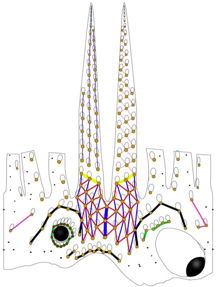
Figure. 360° view of S. hoylei head and arms IV showing photophore arrangement, color coded. Drawing by R. Young.
- Basal Arm IV Row with 3 photophores. Medial photophore separated by 1 photophore from anterior photophore of Midline Series.
- Midline Series with 3 photophores.
- Right Eyelid Series with 17 photophores.
- Left Eyelid Series with 6 photophores.
- 2° Right Eyelid Series with 8 photophores. Photophore no. 8 small and positioned well posterior and dorsal to middorsal point of eyelid.
- 2° Left Eyelid Series with 7 photophores.
- Ventral Matrix with 35 photophores; 9 longitudinal (blue) series; attachment to 2° eyelid series at 2° eyelid photophores no. 2 on either side. Rogue photophore absent.
- Ventral Matrix photophores do not align in transverse rows.
- Right basal series with 2 photophores.
- Left basal series absent.
- Basal row with 8 photophores and 3 sawteeth.
- Right Accessory Series with 2 photophores.
- Left Accessory Series with 3 photophores.
- Simple photophores (round, black dots):
- Arrangement as illustrated in figure.
- Large, simple photophores on dorsal surface of head absent.
- Small, simple photophores not easy to recognize and may be variable.
- Photographs showing photophore patterns can be seen here.
- Compound photophores (Type 1a):
- Compound photophores (mantle)
- Compound photophores of large, uniform size on anterior half of ventral mantle. Click on an image to view larger version & data in a new window
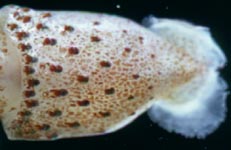
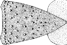
Figure. Left - Ventral view of mantle, small S. hoylei, Hawaiian waters. Photograph by R. Young. Right - Ventral view of mantle, S. hoylei (?), 68 mm ML, 40°S, 125°W. Drawing extracted from Fig 21a of Voss (1969).
- Compound photophores (arms)
- Arms IV with 3 longitudinal series on arm base (photograph below).
- Arms III with one longitudinal series at bases (lower arm in photograph at right).
- Arms II with one longitudinal series at bases (upper arm in photograph at right).
- Arms I with one longitudinal series at bases. Click on an image to view larger version & data in a new window
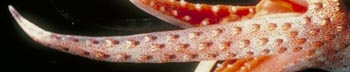
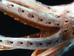
Figure. Left - Ventrolateral view of arm IV, S. hoylei, live squid, Hawaiian waters. Right - Lateral view of left arms II and III, S. hoylei, live squid, Hawaiian waters. Photograph by R. Young.
- Arms
- Length 160-250% of ML. Length arms II-IV in mature male 220-250% of ML, arms I 300-310% of ML (Voss, 1969, composite of all Stigmatoteuthis spp.).
- Arm sucker rings with about 6-16 rounded or truncate teeth on distal and lateral margins; sometimes with numerous, truncate teeth around entire margins on arms IV; in mature males suckers on modified ends of arms I with short, sharply pointed teeth on distal and lateral or entire margins (Voss, et al., 1998, composite of Pacific Ocean Stigmatoteuthis spp.). Click on an image to view larger version & data in a new window
Figure. Oral view of sucker from row 6 arm III, S. hoylei (?), 68 mm ML, 40°S, 125°W. Drawing from Voss (1969, fig. 22c).
- Tentacles
- Club with suckers in 5-7 irregular rows on manus. Median suckers greatly enlarged, more than 4 times size of minute marginal suckers. Click on an image to view larger version & data in a new window
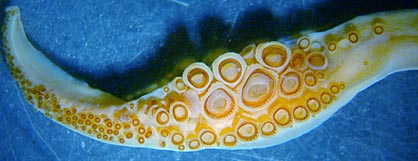
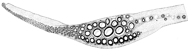
Figure. Oral view of club, S. hoylei, 50 mm ML, Hawaiian waters, preserved. Photograph by R. Young. Oral view of club, S. hoylei (?), 68 mm ML, 40°S, 125°W. Drawing from Voss (1969, fig. 22i).
- Median, enlarged manus suckers with about 55-85 sharp, pointed teeth around entire margins (specimens from 19°N-33°N, 123°-124°W), 38 teeth in a large squid from the South Pacific (40°S, 125°W) (Voss, et al., 1998). Click on an image to view larger version & data in a new window
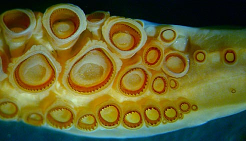
Figure. Oral view of manus suckers, S. hoylei, 50 mm ML, Hawaiian waters, preserved. Photograph by R. Young.
- Club with suckers in 5-7 irregular rows on manus. Median suckers greatly enlarged, more than 4 times size of minute marginal suckers.
- Skin
- Skin covered with low, fleshy pads, giving overall rough appearance in large squid. Click on an image to view larger version & data in a new window
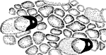
Figure. Ventral view of base of arm IV showing skin warts of S. hoylei (?), 68 mm ML, 40°S, 125°W. Drawing from Voss, 1969 (Fig. 22j).
- Skin covered with low, fleshy pads, giving overall rough appearance in large squid.
- Web
- Inner web virtually absent, outer web 3-8% length of longest arm.
- Inner web virtually absent, outer web 3-8% length of longest arm.
- Reproductive system
- Males with paired genitalia and spermatophore organs.
- Spermatophores
- Length 13.6-16% of ML; sperm mass 59-63% of spermatophore length (SpL); cement body 20-21% of SpL; ejaculatory apparatus 17-19% of SpL with multiple, complex looping (photograph on the right). Connective complex appears to be absent. Click on an image to view larger version & data in a new window
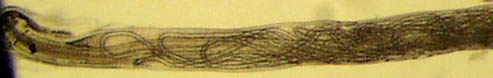
Figure. Oral 3/4 of ejaculatory apparatus of spermatophore of S. hoylei, 100 ML, off Hawaii. Photograph by R. Young.
- Length 13.6-16% of ML; sperm mass 59-63% of spermatophore length (SpL); cement body 20-21% of SpL; ejaculatory apparatus 17-19% of SpL with multiple, complex looping (photograph on the right). Connective complex appears to be absent.
- Hectocotylus
- Both arms I equally modified on distal 2/3. Suckers palisaded on elongate pedestals except near tip. Suckers in two broadly separated series in proximal portion of modified section. Scattered suckers present between marginal series over a middle section of the modified region. Suckers enlarged near tip. Lateral surfaces of arms smooth (no tubercules). More details of the hectocotylus can be found here.
Click on an image to view larger version & data in a new window
Figure. Oral view of portions of both hectocotylized arms I of S. hoylei, 100 mm ML, same specimen as spermatophore. Photograph by R. Young.
Comments
This description, except for the photographs, the head photophores and the description of the hectocotylus, is from Voss (1969) and Voss et al. (1998).


