- Photophores (head)
- Compound photophores (Type 2b):
 Click on an image to view larger version & data in a new window
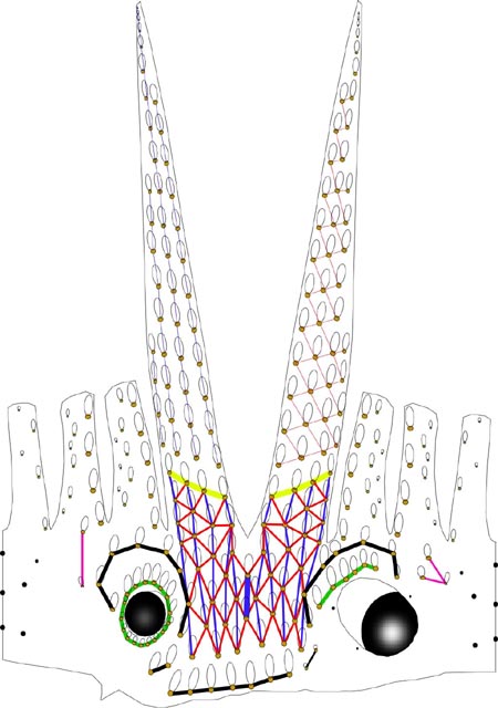
Figure. Arrangement of photophores on head and arms of H. berryi. Dorsal, lateral and ventral views combined in the illustration. The line on either side of the head represents the dorsal midline.
- Basal Arm IV Row with 4 photophores. Medial photophore separated by 2 photophores from anterior photophore of Midline Series.
- Midline Series with 2 photophores.
- Right Eyelid Series with 17 photophores .
- Left Eyelid Series with 6 photophores.
- 2° Right Eyelid Series with 8 photophores.
- 2° Left Eyelid Series with 7 photophores.
- Ventral Matrix with 48 photophores; 11 longitudinal (blue) series; attachment to 2° Eyelid Series at 2° eyelid photophores no. 2 on either side.
- Ventral Matrix photophores tend to align in transverse rows.
- Right basal series with 2 photophores.
- Left basal series with two very small photophores.
- Basal row smooth alignment with 7 photophores .
- Right Accessory Series with 2 photophores (see below).
- Left Accessory Series with 3 photophores (see below).
Simple photophores (round, black dots):
- Arrangement as illustrated.
- Large, simple photophores on dorsal surface of head.
- Small, simple photophores not easy to recognize and may be variable.
Photographs showing arrangement of photophores can be found here.
- Compound photophores (Type 2b):
- Mantle Photophores
- Compound photophores of uniform size in anterior half of ventral mantle.
Click on an image to view larger version & data in a new window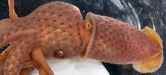
Figure. Ventral view of mantle of H. berryi, preserved specimen. Photograph by R. Young.
- Arm Photophores
- Arms IV without gap between proximal arm photophores and arm tip photophores (photograph above).
- Arms IV with photophores in four photophore series at base (photograph above).
- Arms III with two major longitudinal photophore series at bases (photograph below); one minor photophore series present (see drawing at top of page).
- Arms II with two major longitudinal photophore series at bases (photograph below); one minor photophore series seems to blend with dorsal major series (see drawing at top of page).
- Arms I with one major longitudinal photophore series at bases; one minor photophore series present (see drawing at top of page).
Click on an image to view larger version & data in a new window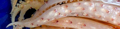

Figure. Left - Ventral view of arm IV of H. berryi, 49 mm ML, 31°N, 155°W. Right - Lateral view of arms II (top) and III, same specimen. Photograph by R. Young.
- Arms and Web
- Inner web moderately deep, ca. 15-25% of longest arm.
- Length 95-120% of ML
- Suckers of ventral-marginal series with similar dentition (right, bottom photograph).
- Arm I-III suckers with smooth inner rings (photograph below, left) except at tips where toothed suckers are present (photograph below, middle).
- Sucker rings of arms IV with numerous, square, tightly=packed teeth on entire or distal margins (photograph below, right).
Click on an image to view larger version & data in a new window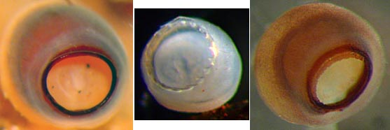
Figure. Oral views of arm suckers of H. berryi, preserved. Left - Oral view of arm I sucker, 48 mm ML, Hawaiian waters. Middle - Arm-tip suckers of arm III, 49 mm ML, 31°N, 155°W. Right - Arm IV sucker, outer ring removed, 48 mm ML, Hawaii waters. Photographs by R. Young.
- Tentacles
- Approximately 5 irregular sucker series on club.
- Central manus suckers 2 x diameter of marginal suckers.
- Sucker rings on enlarged suckers of the manus with 28-34 triangular teeth around entire margin (right, top photograph).
- Buccal crown
- Buccal crown with 7 supports.
Click on an image to view larger version & data in a new window
Figure. Oral view of buccal crown of H. berryi, holotype. Drawing from Voss, 1969 (Fig. 17f).
- Funnel
- Dorsal pad of funnel organ swollen and without sculpture.
Click on an image to view larger version & data in a new window
Figure. Ventral view of funnel organ of H. berryi, holotype. Drawing from Voss, 1969 (Fig. 17e).
- Fins
- Length 40-44% of ML, width 56-65% of ML.
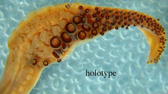
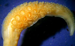
Figure. Oral view of tentacular clubs of H. berryi. Left - holotype. Right - 49 mm ML, 49 mm ML, 31°N, 155°W. Photographs by R. Young.
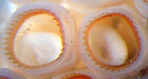
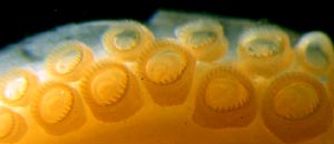
Figure. Oral view of club suckers of H. berryi, 49 mm ML, 31°N, 155°W. Left - Large club suckers. Right - Marginal club suckers, same specimen. Photographs by R. Young.
Comments
Except for the information on photophores of the head and the photographs, the above data comes from Voss (1969)


