
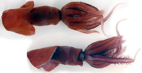
Figure. P. sloani. Left - Dorsal and lateral views of the holotype, female, 58 mm ML. Right - Dorsal view of the paratype, female, 102 mm ML.
- Arms
- Arms thick, without keels and with low, thick protective membranes.
- Arm suckers with smooth inner rings having the appearance of fused, truncated teeth.
- Tentacles
- Club suckers small; few scattered suckers at club base become much more numerous distally.
- Club suckers on white stalks emerging from small, white circular patches on pigmented club; papillae between suckers absent.
- Club diameter narrows somewhat more abruptly where suckers become more densely packed, forming a nearly cylindrical distal end.
- Club gradually encircles increasing portion of tentacle distally to a maximum of ca. 2/3s of tentacle circumference.
- Dark ridge and papillae series do not extend to tip of club.
- Club suckers with small, irregular, knob-like teeth aroung entire margin on inner ring.
- Buccal Crown
- Ventral buccal connectives attach to ventral margins of arms IV.
- Head
- Head without detectable olfactory organ; without occipital folds; without distinct funnel groove; without visible funnel bridles; without canal between bridles.
- Beaks
- Lower beak with short rostrum; with convex jaw edge; without lateral wall fold or ridge.
- Upper beak with short rostrum; with hood margin high above lateral wall crest.
- Funnel
- Funnel locking-apparatus with oval depression.
- Fins
- Mantle extends to posterior end of fins.
- Photophores
- Photophores absent.
- Pigmentation
- General pigmentation purple-brown. Pigment in epithelial cells; chromatophores appear to be absent.
- Light to absent pigmentation over eye lenses, aboral base of tentacles and arms IV, distal ends of tentacles; white line down aboral midline of tentacles over proximal 60% of tentacle length.
- Surfaces within the mantle cavity heavily pigmented.
- Gladius
- Gladius of the paratype according to Toll (1982):"A stiff, dense, gelatinous material fills the ventral concavity of the rachis and vanes. It is bilobed venrally in the two expanded portions of the vanes, domed in the narrow portion."
- Measurements of paratype gladius from Toll (1982): Gladius length - 93.5 mm. Anterior gladius width - 7.4 mm. Posterior gladius width - 10.3 mm. Free rhachis length - 26 mm. Free rhachis width - 1.9 mm.
- Viscera
- Anus without anal valves.
- Measurements
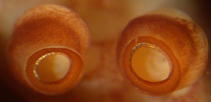
Figure. Oral views of large suckers from arm II of P. sloani, holotype, preserved.
Scanning electron micrographs of the suckers of the paratype can be seen here.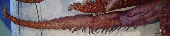
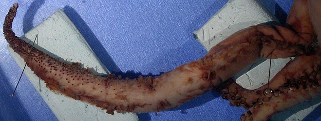
Figure. Top - Oral view of the right tentacle of P. sloani, holotype, preserved. Note the shape change near the level of the right insect pin. Bottom - Oral-lateral view of the tentacle, paratype, preserved. Note the difference in the shape of the papillae.

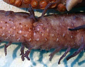
Figure. Left - Aboral view of the club of P. sloani near the tip, holotype, preserved. Arrows point to a diminishing pigmented ridge (top)and a papilla-series (bottom). Note that the aboral surface is virtually bare to the club tip. Right - Oral view of base of club, holotype, preserved. Note the branches on the papillae and the unpigmented patches from which the sucker stalks arise.
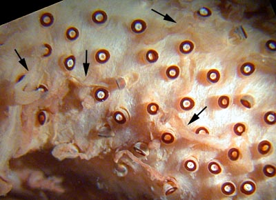
Figure. Oral view near the proximal end of the club of P. sloani, paratype, showing long, slender, simple papillae interspersed with the suckers. These papillae were not seen on the holotype.
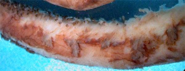

Figure. Views of the club papillae of P. sloani, paratype. Top - Lateral view of the basal portion of the club. Bottom - Aboral view of the club. Unlike the more simple papillae of the holotype, the papillae of the paratype are flattened and have highly complex branching patterns.
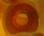
Figure. Oral view of large club suckers (0.2 mm across outer ring)of P. sloani, holotype, preserved.
Scanning electron micrographs of the club suckers of the paratype can be seen here.A dissection of the peculiar eye of the paratype can be seen here.
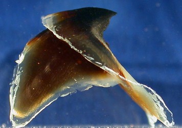
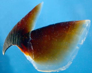
Figure. Side views of beaks of P. sloani, paratype. Left - Lower beak. Right - Upper beak.
More photographs of the beaks can be found here.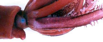
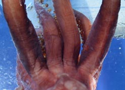
Figure. Pigmentation of P. sloani, holotype. Left - Side view brachial crown showing aboral surfaces of tentacle bases, unpreserved. Right - Ventral view of brachial crown showing aboral surfaces of tentacle bases, preserved. Light line present down aboral surface of tentacles and slight area of light pigmentation present at aboral tentacle base and arm IV bases
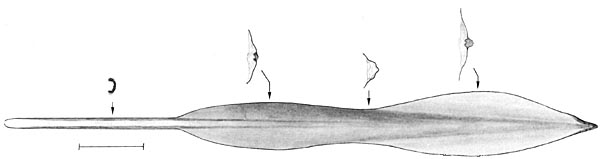
Figure. Ventral view of the gladius of P. sloani, paratype, with cross-sections above. Arrows indicate locations of cross-sections. Drawing from Toll (1982).
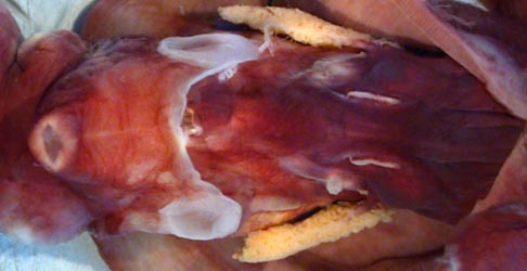
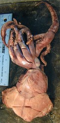
Figure. Ventral view of the mantle cavity of P. sloani with mantle cut open and reflected backward, holotype, preserved. The pair of thin, white lines are the nidamental glands. Renal papillae are also visible. Anus is visible just posterior to the funnel. Funnel locking-apparatus does not show normal shape and the delicate structure was flattened in preservation.
| Holotype | Paratype | |
| Sex | Immature female | Immature female |
| Mantle length | 58 | 102 present; 112 (Toll, 1982) |
| Gladius length | 93.5* | |
| Mantle width | Ca. 18 | - |
| Head width | 13 | - |
| Head length* | 6.5 | 34 |
| Fin length | 33 | 70 |
| Fin width | 54 | 121 |
| Arm I, length, left/right | 31 / 32 | 83+ / 92 |
| Arm II, length | 35 / 37 | - / 93 |
| Arm III, length | 32 / 29 | - / 88 |
| Arm IV, length | 35 / 34 | - / 90 |
| Tentacle length | 60 / 60 | 148 / - |
| Club length | 36 / 37 | 71 / - |


