Drawings below are first made from photographs. The drawings should then be compared, under a microscope, with the preserved squid from which the photographs were made. This greatly reduces the errors that can occur in interpreting the photophore distributions. Use caution, therefore, on this page in viewing the color and distribution of the dots (representing photophores) as their distributions have not yet been confirmed by comparing the drawings against the actual specimen under the microscope.

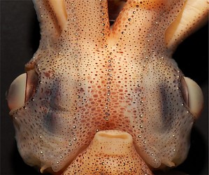
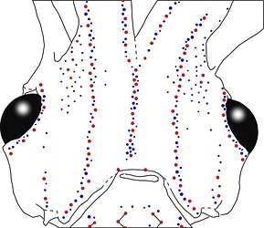
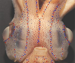
Figure. Ventral views of the integumental photophores of A. affinis, mature male, 33 mm ML. Left - Photograph of preserved squid. Middle - Outline drawing from photograph with all integumental photophores represented by colored dots. Right - Photograph with superimposed dots on integumental photophores. Red dots - Complex photophores. Blue dots - Non-complex photophores. Images by R. Young.
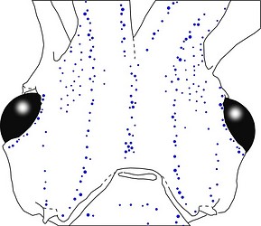
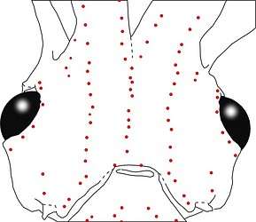
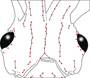
Figure. Ventral views of the integumental photophores of the same A. affinis. Left - Drawing showing only the non-complex (blue) photophores. Middle - Drawing showing only the complex (red) photophores . Right - Same drawing with lines connecting red photophores to aid comparisons of photophore patterns between species. Images by R. Young.
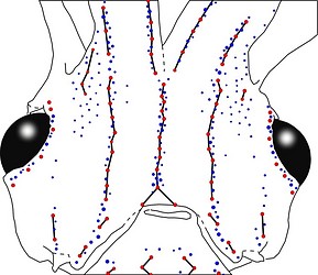
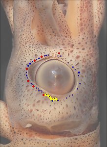
Figure. Left - Same squid, same view. Drawing showing all photophores and lines showing red photophore patterns. Middle - Side view of the same preserved squid showing photophores of the eyelid. Right - Same photograph (dimmed) with red and blue dots superimposed on photophores. Yellow line connects photophores of the Posterior Eyelid Series. Images by R. Young.
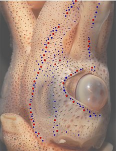
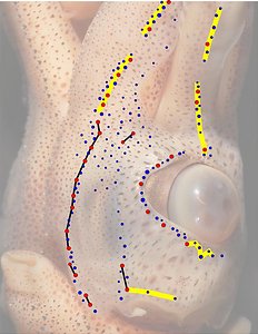
Figure. Oblique (ventrolateral) view of the same squid showing the lateral photophores. Left - Photograph of the preserved squid. Middle - Same photograph, dimmed, with red and blue dots superimposed on photophores to more clearly show the distribution of the two photophore types. Right - Same photograph, dimmed further, with black lines connecting some "red" photophores to assist in comparing red-photophore patterns and yellow lines connecting some blue-red-photophore patterns. The latter combines both types of photophores to define certain patterns. Both types can be important in comparing patterns between species. Images by R. Young.
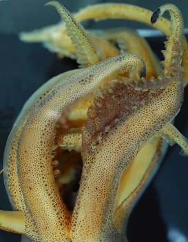
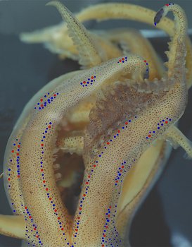
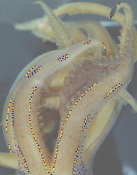
Figure. Ventral view of A. affinis, mature male, ? mm ML. Left - Composite photograph of the arms of the preserved squid. Middle - Same photograph, dimmed, with all integumental photophores represented by colored dots. Right - Same photograph, dimmed further, with yellow lines connecting blue-red-photophore patterns. The discontinuous arrangement of photophores on the arms are best defined by the combination of both types of photophores. Images by R. Young.
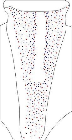
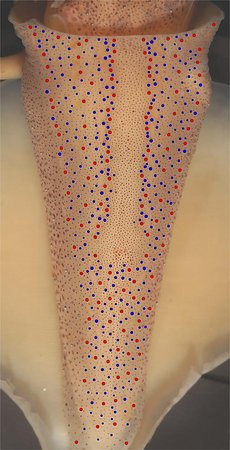
Figure. Ventral views of the integumental photophores of the mantle of A. affinis, mature male, 29 mm ML, Eastern Tropical Pacific. Left - Photograph of the preserved squid. Middle - Outline drawing from the photograph with all integumental photophores represented by colored dots. Right - Photograph with dots superimposed dots on integumental photophores. Red dots - Complex photophores. Blue dots - Non-complex photophores. Images by R. Young.
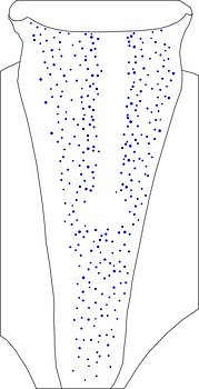
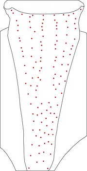
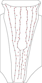
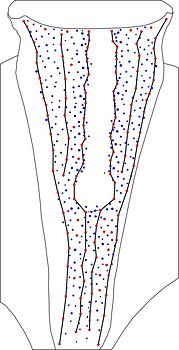
Figure. Integumental photophores of the mantle continued. Left - Drawing showing only the non-complex (blue) photophores. Left middle - Drawing showing only the complex (red) photophores. Right middle - Same drawing with lines connecting red photophores to aid comparisons of photophore patterns between species. Note that all red photophores do not fall on the lines. Right - Drawing showing all photophores and lines showing red photophore patterns. Images by R. Young.
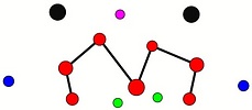
Figure. Ventral view of the funnel-groove region of A. affinis, mature male, 26 mm ML. Top - Funnel groove showing photophores of funnel groove and surrounding tissue. Middle - In the photograph, white dots are placed over the "white" photophores of the funnel groove. Black dots are placed over the two most posteromedial "red" photophores of the ventral head to mark the anterolateral edges of the funnel groove. Bottom - Same pattern of dots as in photographs but dots are colored. Colored dots, lines - Aid in comparison of patterns between species. Images by R. Young. Comparison of white photophore patterns for all Abraliopsis species can be found here.
Comments 1: Summary of photophore distributions. (photophore definitions found here)
ARMS:
Medial Arm IV Series - Continuous; reaches about half of arm length.
Central Arm IV Sector - Photophores absent.
Lateral Arm IV Series - Discontinuous; long gaps separate patches; extends to arm tip.
Lateral Membrane Series of arms IV - Reaches about half of arm length.
Arm III Series - Discontinuous; long gaps separate patches, extends nearly to arm tip.
HEAD - Typical three photophore series on central-ventral head with:
Median Head Series - 3 "red" photophores in posterior caret. Anteriorly red photophores in irregular single series. Blue photophores absent from within the triangle limited by the caret, and, mostly, including lines that form the caret.
Median Head Sector - Absent.
Lateral Head Sector - Without photophores except for a few blue photophores in the anterolateral area of this sector in large squid.
Lateral Head Series - Single, nearly straight, series of red photophores.
Window Sector - Many, scattered, blue photophores anterolaterally, otherwise photophores absent.
1st Lateral Window Series - Two segments broadly separated by mostly single series of blue photophores: posterior segment with 2 red photophores; anterior segment with two, closely spaced, or one (one side only, 25 mm ML) red photophore. One squid appears to have two additional red photophores extending onto lateral arm IV membrane but only on the right side.
2nd Lateral Window Series - Absent.
Eyelid Series - Red photophores only on ventral half of eyelid. Posterior Eyelid Series with two large blue photophores displaced slightly posteriorly from eyelid series; more ventral of two, larger and more posterior. Formula: RbbBbB.
Occipital Series - Rbb.
Lateral Funnel-groove Series - 4 red photophores, in two segments not aligned.
White patch (funnel groove) - Complex-2 pattern (red, green, blue; + maroon at larger ML, + yellow ?).
FUNNEL:
Medial Patch - "Nose" pattern of 4 "red" photophores, but indication that some variation may also occur.
Lateral Patch - Two red photophores.
MANTLE - Typical 6 photophore series on ventral mantle with:
Median Mantle Sector - Broad and without photophores (bare) in mid-region in anterior half of mantle. At its posterior end this sector expands laterally. The result is a sector with a key-hole shape. Further posteriorly the sector narrows greatly and is virtually obscure due to scattererd blue photophores. Blue photophores in the lateral regions of this key-hole portion of this sector lie unusually close to the the medial mantle series of red photophores. That is, this key-hole region has few blue photophores and these are found only at its periphery.
Medial Mantle series - Somewhat irregular alignment in anterior half of mantle.
1st Lateral Mantle Sector - Numerous scattered blue photophores more concentrated along medial margins of sector in anterior half of mantle. One to four "free" red photophores (i.e., not part of a standard series). Sector cannot be followed beyond first half of mantle.
2nd Lateral Mantle sector - Numerous blue photophores but less abundant than in First sector. One "free" red photophore near anterior mantle margin. Sector cannot be followed beyond first half of mantle.
Lateral Mantle Series - Posteriorly, scattered "free" red photophores that cannot clearly be assigned to any Series.
Mantle-angle Series - Fairly straight alignment (alignment anteriorly veers laterally as mantle broadens).
Comments 2: Unusual features of the photophore patterns.
- Lateral arm IV series and arm III series are discontinuous with (1) long gaps between photophore patches and (2) gaps begin near proximal end of arms.
- Caret at posterior end of Median Head Series with (1) 3 red photophores and (2) mostly without blue photphores on or within caret.
- Segments of First Lateral Window Series very broadly separated. Needs more study, however, to determine variability.
- Mantle with (1) key-hole shaped portion of median mantle sector in anterior half of mantle; with (2) few blue photophores within key-hole portion, most positioned tight to lateral margins (see last mantle illustration above) and (3) some "free" red photophores in Mediolateral Mantle Sectors.


