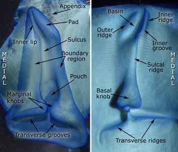
The ommastrephid funnel/mantle locking-apparatus is one of the most characteristic features of the family. The funnel component is cartilagenous and contains a longitudinal groove (sulcus) that is swollen posteriorly (pouch) and tapers anteriorly, and posterior transverse grooves. The sulcus is surrounded by the boundary region which may be modified ito form a lip and pad anteriorly. The medial wall of the sulcus is more clearly defined than the lateral wall and generally undercuts the adjacent boundary region for much of its length. Some of the cartilage extends into the funnel muscle to form an anchor and a potion of the anchor extends anteriorly beyond the lock just beneath the integument (appendix). The mantle component has a fibrous construction except anteriorly where some cartilage overlies part of its ridges and grooves.

Ommastrephinae
- Ommastrephes bartramii
- Funnel component
- Anterior end strongly asymmetrical.
- Anterior Sulcus narrow and sharply bent medially; bordered medially by slender Inner Lip and laterally by broad Pad.
- Pad with abrupt posterior termination at bend in Sulcus; grades into lateral wall of Sulcus.
- Large, cartilagenous, triangular Appendix present.
- Anterior cross-section (medial-lateral): narrow Inner Lip, narrow Sulcus, Outer Lip (sharp inner edge of the pad), Pad.
- Anterior end strongly asymmetrical.
- Mantle component
- Anterior end with abrupt medially bend in slender Sulcal Ridge.
- Anterior Sulcal Ridge bordered medially by narrow Inner Groove (the inner lip inserts here) and laterally by broad Basin (the Pad inserts here) bound by Outer Ridge, giving tip bifurcate appearance.
- Lateral side of Sulcal Ridge with broad taper that merges into Basin; medial side of Ridge sharply defined.
- Cartilagenous tip covers much of slender Sulcal Ridge and medial portion of Basin; small anteromedial cartilagenous extension covers tip of Medial Ridge.
- Anterior cross-section (medial-lateral): slender Inner Ridge, narrow Inner Groove, slender Sulcal Ridge, Basin, Outer Ridge (outer wall of basin).
Click on an image to view larger version & data in a new window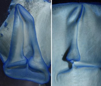
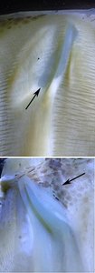
Figure. Left - Frontal views of the funnel and mantle components of the funnel locking-apparatus of Ommastrephes bartramii, immature male, 155 mm ML, Hawaiian waters, stained with methylene blue stain. Right - Anterior ends of the mantle (top) and funnel (bottom) components, partially destained. Arrows point to cartilages. Triangular cartilagenous appendix just visible through skin. Photographs by R. Young.
Click on an image to view larger version & data in a new window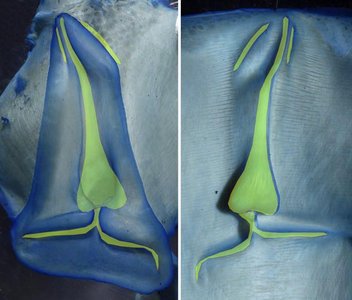
Figure. Same funnel and mantle components of the funnel locking-apparatus of Ommastrephes bartramii as seen above. The yellow color on the mantle component highlights ridges. These highlights have then been reversed in orientation and overlaid on the funnel component to show how the two components combine. Photographs by R. Young.
- Funnel component
- Sthenoteuthis oualaniensis
- Funnel component
- Anterior end strongly asymmetrical.
- Anterior Sulcus narrow and sharply bent medially; bordered medially by slender Inner Lip and laterally by broad Pad.
- Pad with abrupt posterior termination at bend in Sulcus.
- Anterior cross-section (medial-lateral): narrow Inner Lip, narrow Sulcus, Outer Lip (sharp inner edge of the pad), Pad.
- Large, cartilagenous, triangular Appendix present.
- Posterior portion of Sulcus and medial Transverse Groove fused to mantle component via fibrous connection.
- Mantle component
- Anterior end with abrupt medially bend in slender Sulcal ridge.
- Anterior Sulcal Ridge bordered medially by narrow Inner Groove (the inner lip inserts here) and laterally by broad basin (the pad inserts here) bound by Outer Ridge, giving tip bifurcate appearance..
- Lateral side of Sulcal Ridge with moderate taper that ends posterior to the Basin in a low dome.
- Anterior cross-section (medial-lateral): slender inner ridge, narrow inner groove, sulcal ridge, basin, outer ridge (outer wall of basin).
- Posterior portion of Sulcal Ridge and medial Transverse Ridge fused to mantle component via fibrous connection.
- Cartilagenous tip covers all of narrow sulcal ridge and most of posterior portion of basin; broad anteromedial cartilagenous extension covers medial ridge.
- Posterior portion of Sulcal Ridge and medial Transverse Ridge fused to mantle component via fibrous connection.
Click on an image to view larger version & data in a new window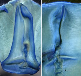
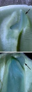
Figure. Left - Frontal views of the funnel and mantle components of the funnel locking-apparatus of Sthenoteuthis oualaniensis, immature female, 140 mm ML, Hawaiian waters, stained with methylene blue stain. Note the tissue cut to separate the fused components. The arrow points to a strip of tissue torn out of the mantle when the components were pulled apart but the fusion hadn't been completely cut. Right - Anterior ends of the mantle (top) and funnel (bottom) components, partially destained. Arrows point to cartilages. Photographs by R. Young.
- Funnel component
- Sthenoteuthis pteropus
- As in S. oualaniensis except funnel and mantle components not fused.
- As in S. oualaniensis except funnel and mantle components not fused.
- Dosidicus gigas
- Funnel component
- As in Ommastrephes bartramii.
- As in Ommastrephes bartramii.
- Mantle component
- As in Ommastrephes bartramii except midregion of Sulcal Ridge appears nearly flattened in our specimen. We suspect that this is not normal but additional material is needed.
- Anterior cartilagenous tip less extensive than in O. bartramii but still has 3-part components: posterior Basin, anterior narrow Sulcal Ridge, anterior tip of Inner Ridge.
Click on an image to view larger version & data in a new window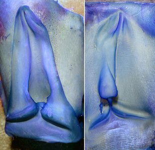
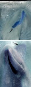
Figure. Left - Frontal views of the funnel and mantle components of the funnel locking-apparatus of Dosidicus gigas, mature male, 186 mm ML, near Galapagos Islands, stained with methylene blue stain. Right - Anterior ends of the mantle (top) and funnel (bottom) components, partially destained. Arrows point to cartilages. Skin removed over Appendix. Photographs by R. Young.
- Funnel component
- Eucleoteuthis
- Funnel component
- Anterior end with slight, asymmetrical appearance due to broader lateral than medial Boundary Region.
- Distal sulcus narrow; Inner Lip and laterally by Pad more poorly defined than in O. bartamii.
- Pad represents slightly modified anterior portion of lateral Boundary Region.
- Anterior cross-section (medial-lateral): Inner Lip, Sulcus, narrow Pad.
- Large, cartilagenous, triangular Appendix present.
- Posterior portion of sulcus and medial transverse groove fused to mantle component via fibrous connection.
- Mantle component
- Anterior end with snearly straight Sulcal Ridge.
- Distal sulcal ridge bordered medially by inner groove and laterally by narrow basin bound by Outer Ridge, giving tip bifurcate appearance.
- Lateral side of Sulcal Ridge with broad taper that merges into Basin; medial side of Ridge sharply defined.
- Anterior cross-section (medial-lateral): Inner Ridge, Inner Groove, Sulcal Ridge, slender Basin, outer Ridge.
- Cartilagenous tip covers distal half of narrowed Sulcal Ridge and midportion of Basin; anteromedial cartilagenous extension covers tip of Medial Ridge.
- Posterior portion of Sulcal Ridge and medial Transverse Ridge fused to mantle component via fibrous connection.
Click on an image to view larger version & data in a new window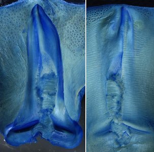
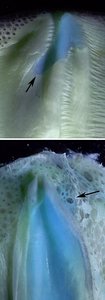
Figure. Left - Frontal views of the funnel and mantle components of the funnel locking-apparatus of Eucleoteutis luminosa, mature female, 180 mm ML, northern Hawaiian waters, stained with methylene blue stain. Note the tissue cut to separate the fused components. Right - Anterior ends of the mantle (top) and funnel (bottom) components, partially destained. Arrows point to cartilages. Photographs by R. Young.
- Funnel component
- Hyaloteuthis pelagica
- Funnel component
- Anterior end nearly symmetrical but with Boundary Region distinctly broader on lateral side, without lateral bend.
- Narrow anterior Sulcus without distinct Inner Lip or lateral Pad.
- Sulcus nearly straight.
- Anterior cross-section (medial-lateral): Boundary Region, Sulcus, Boundary Region.
- Moderately large, triangular cartilagenous Appendix present.
- Mantle component
- Sulcal Ridge nearly straight.
- Anterior Sulcal Ridge bordered medially by Inner Groove and Inner Ridge and laterally by somewhat broader groove (Basin) and Outer Ridge, giving tip subtle, bifurcate appearance.
- Anterior cross-section (medial-lateral): Inner Ridge, Inner Groove, Sulcal Ridge, outer groove, Outer Ridge.
- Cartilagenous tip covers tip of Sulcal Ridge, portion of outer groove and tip of Medial Ridge.
- Anterior cross-section (medial-lateral): broad Inner Ridge, narrow Inner Groove, slender Sulcal Ridge, broad outer groove, Outer Ridge.
Click on an image to view larger version & data in a new window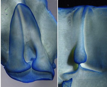
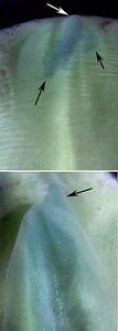
Figure. Left - Frontal views of the funnel (58 mm ML, mature female) and mantle (51 mm ML, male, probably mature) components of the funnel locking-apparatus of Hyaloteuthis pelagica, Hawaiian waters, stained with methylene blue stain. Right - Anterior ends of the mantle (top) and funnel (bottom) components, partially destained. Arrows point to cartilages. Arrow points to cartilage. Photographs by R. Young.
- Funnel component
- Ornithoteuthis antillarum
- Funnel component
- Anterior end virtually symmetrical.
- Sulcus narrows distally with a slight medial curve.
- Sulcus with lateral edge weakly defined in mid-region.
- Small, pointed Appendix present.
- Anterior cross-section (medial-lateral): medial Boundary Region, Sulcus, lateral Boundary Region.
- Mantle component
- Sulcal Ridge narrows distally with a slight medial curve.
- Anterior Sulcal Ridge bordered on either side by broad swelling of muscular mantle wall. Swellings may be artifact or could be homologous with Outer and Inner Ridges of O. bartramii and similar species.
- Lateral side of middle Sulcal Ridge with broad taper; medial side of Ridge sharply defined.
- Cartilagenous tip covers much of slender, anterior Sulcal Ridge with slight flare along either side.
- Anterior cross-section (medial-lateral): swelling, Sulcal Ridge, swelling.
Click on an image to view larger version & data in a new window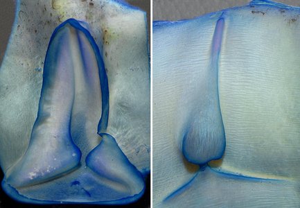
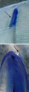
Figure. Left - Frontal views of the funnel and mantle components of the funnel locking-apparatus of Ornithoteuthis antillarum, mature female, 91 mm ML, equatorial Atlantic, stained with methylene blue stain. Right - Anterior ends of the mantle (top) and funnel (bottom) components, partially destained. Cartilagenous appendix (at tip of arrow) photographed after skin removed. Photographs by R. Young.
- Funnel component
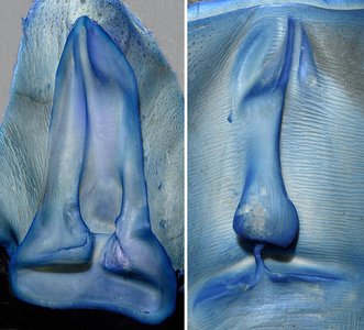
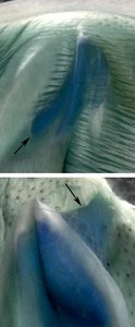
Figure. Left - Frontal views of the funnel and mantle components of the funnel locking-apparatus of Sthenoteuthis pteropus, immature female, 165 mm ML, Gulf of Mexico, stained with methylene blue stain. Marginal and basal knobs slightly damaged during preparation. Right - Anterior ends of the mantle (top) and funnel (bottom) components, partially destained. Arrows point to cartilages. Photographs by R. Young.
Illicinae
- Illex illecebrosus
- Funnel component
- Anterior end virtually symmetrical.
- Sulcus narrows distally but remains virtually straight.
- Sulcus with medial and lateral edges approximately equally well defined.
- Large, rounded cartilagenous Appendix present.
- Anterior cross-section (medial-lateral): medial Boundary Region, Sulcus, lateral Boundary Region.
- Mantle component
- Sulcal Ridge narrows distally but remains virtually straight.
- Anterior Sulcal Ridge bordered on either side by broad swelling of muscular mantle wall. Swellings may be artifact or could be homologous with Outer and Inner Ridges of O. bartramii and similar species.
- Sulcal Ridge with medial and lateral edges approximately equally well defined.
- Cartilagenous tip covers much of slender, anterior Sulcal Ridge with slight flare along either side.
- Anterior cross-section (medial-lateral): slight swelling, Sulcal Ridge, slight swelling.
Click on an image to view larger version & data in a new window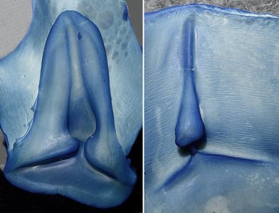
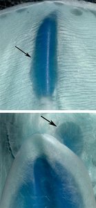
Figure. Left - Frontal views of the funnel and mantle components of the funnel locking-apparatus of Illex illecebrosus, mature female, 134 mm ML, western North Atlantic, stained with methylene blue stain. Right - Anterior ends of the mantle (top) and funnel (bottom) components, partially destained. Arrows point to cartilages. Skin removed over Appendix. Photographs by R. Young.
- Funnel component
Todarodinae
- Todarodes pacificus
- Funnel component
- Anterior end nearly symmetrical.
- Sulcus narrows distally but with distinct bend.
- Sulcus with lateral edge weakly defined in mid-region.
- Moderate-sized, triangular, cartilagenous Appendix present.
- Anterior cross-section (medial-lateral): medial Boundary Region, Sulcus, lateral Boundary Region.
- Mantle component
- Sulcal Ridge narrows distally but without distinct bend.
- Anterior Sulcal Ridge bordered on either side by narrow swelling of muscular mantle wall. Swellings may be artifact or could be homologous with Outer and Inner Ridges of O. bartramii and similar species.
- Lateral side of middle Sulcal Ridge with broad taper; medial side of Ridge sharply defined.
- Cartilagenous tip covers much of slender, anterior Sulcal Ridge and much of posterolateral region up outer swelling.
- Anterior cross-section (medial-lateral): narrow inner swelling, broad inner depression, slender Sulcal Ridge, broad outer depression, narrow outer swelling.
Click on an image to view larger version & data in a new window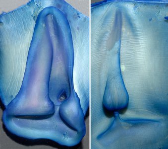

Figure. Left - Frontal views of the funnel and mantle components of the funnel locking-apparatus of Todarodes pacificus, immature male, 214 mm ML, Japanese waters, stained with methylene blue stain. The hole in the pouch is due to damage during preparation. Right - Anterior ends of the mantle (top) and funnel (bottom) components, partially destained. Arrows point to cartilages. Skin removed over Appendix. Photographs by R. Young.
- Funnel component
- Todaropsis eblanae
- Funnel component
- Anterior end nearly symmetrical.
- Sulcus very short, narrows distally but Sulcus and Pouch form straight line that strongly tilts medially.
- Sulcus with lateral edge only slightly more weakly medial edge.
- Presence of Appendix undetermined.
- Anterior cross-section (medial-lateral): medial Boundary Region, Sulcus, lateral Boundary Region.
- Mantle component
- Sulcal Ridge very short, narrows distally but Sulcal Ridge and Pouch form straight line that strongly tilts medially.
- Anterior Sulcal Ridge bordered on either side by swelling of muscular mantle wall. Swellings may be artifact or could be homologous with Outer and Inner Ridges of O. bartramii and similar species.
- Sulcal Ridge with lateral edge only slightly more weakly medial edge.
- Cartilagenous tip undetermined.
- Anterior cross-section (medial-lateral): swelling, Sulcal Ridge, swelling.
Click on an image to view larger version & data in a new window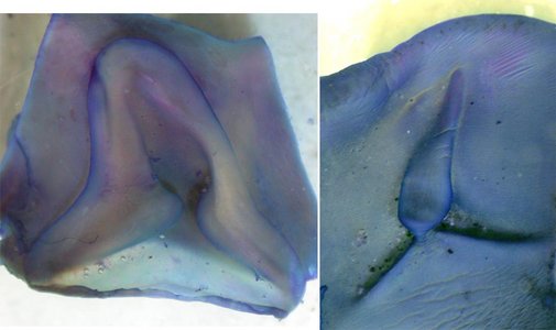
Figure. Frontal views of the funnel and mantle components of the funnel locking-apparatus of Todaropsis eblanae, immature female, 76 mm ML, equatorial Atlantic, stained with methylene blue stain. Photographs by M. Vecchione.
- Funnel component
- Nototodarus hawaiiensis
- Funnel component
- Anterior end nearly symmetrical.
- Sulcus narrows distally with distinct bend.
- Sulcus with lateral edge slightly more weakly defined in mid-region.
- Cartilagenous appendix barely visible from frontal view, extends mostly dorsally into muscle.
- Anterior cross-section (medial-lateral): Boundary Region, Sulcus, Boundary Region.
- Mantle component
- Sulcal ridge narrows distally, with gentle medial curve.
- Anterior Sulcal Ridge bordered on either side by swelling of muscular mantle wall. Swellings may be artifact or could be homologous with Outer and Inner Ridges of O. bartramii and similar species.
- Lateral side of middle Sulcal Ridge with steep taper; medial side of Ridge sharply defined.
- Cartilagenous tip covers much of slender, anterior Sulcal Ridge with slight flare along either side.
- Anterior cross-section (medial-lateral): swelling, narrow Sulcal Ridge, swelling.
- Sulcal ridge ends well posterior to mantle margin; approaches margin in small specimens.
Click on an image to view larger version & data in a new window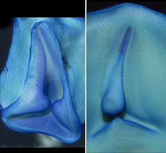
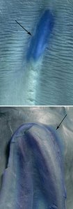
Figure. Left - Frontal views of the funnel and mantle components of the funnel locking-apparatus of Nototodarus hawaiiensis, female, 135 mm ML, Hawaiian waters, stained with methylene blue stain. Right - Anterior ends of the mantle (top) and funnel (bottom) components, partially destained. Cartilagenous appendix (at tip of arrow) photographed after skin removed; appendix short and deeply burried, edges difficult to determine. Photographs by R. Young.
- Funnel component
- Martialia hyadesi *specimen in poor shape
- Funnel component
- Anterior end nearly symmetrical.
- Sulcus narrows distally but with distinct bend.
- Sulcus with lateral edge slightly more weakly defined in mid-region.
- Small-sized, triangular, cartilagenous Appendix present.
- Anterior cross-section (medial-lateral): medial Boundary Region, Sulcus, lateral Boundary Region.
- Mantle component
- Sulcal Ridge narrows distally, with gentle curve.
- Anterior Sulcal Ridge bordered on either side by narrow swelling of muscular mantle wall. Swellings may be artifact or could be homologous with Outer and Inner Ridges of O. bartramii and similar species.
- Lateral side of middle Sulcal Ridge with steep taper; medial side of Ridge sharply defined.
- Cartilagenous tip covers much of slender, anterior Sulcal Ridge and extends well into area between Sulcal Rdige and outer swelling.
- Anterior cross-section (medial-lateral): broad inner swelling, narrow inner depression, slender Sulcal Ridge, broad outer depression, narrow outer swelling.
Click on an image to view larger version & data in a new window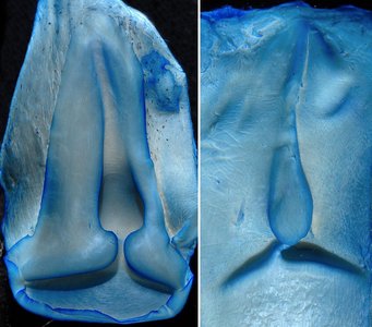
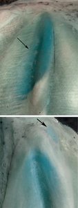
Figure. Left - Frontal views of the funnel and mantle components of the funnel locking-apparatus of Martialia hyadesi, immature female, 180 mm ML, western South Atlantic, stained with methylene blue stain. Right - Anterior ends of the mantle (top) and funnel (bottom) components, partially destained. Arrows point to cartilages. Skin removed over Appendix. Photographs by R. Young.
- Funnel component
Comments
This first look at the comparison of the locking-apparatus of all genera of Ommastrephidae lacks knowledge of the variation at intraspecific and intrageneric levels. The differences seen here, therefore, must be used cautiously.
The higher ommastrephinae (Dosidicus, Sthenoteuthis, Ommastrephes, Eucleoteuthis and Hyaloteuthis) all exhibit considerable similarities in their locks. All have mantle components with bifurcate anterior ends or their remnants and a tripartite distal cartilage. Dosidicus, Sthenoteuthis and Ommastrephes each have a strongly asymmetrical anterior end of the funnel component. The most divergent of these five genera is Hyaloteuthis which may be a result of its small size. Ornithoteuthis, Illex and Nototodarus all have simple locks. Illex appears to be distinguished by the near lack of asymmetry between the medial and lateral sides of the Sulcus and Sulcal Ridge. Todarodes and Martialia share similarities in the anterior cartilage of their mantle components. Todaropsis is distinctive in its short Sulcus and Sulcal Ridge. Both Todaropsis and Nototodarus have the mantle component terminating anteriorly well short of the anterior mantle edge. Also there is considerable differences in the size and shape of the cartilagenous Appendix in some genera.


