- Arms
- Large arm suckers with 10-15 pointed to truncate teeth on distal and lateral margins of inner ring.
 Click on an image to view larger version & data in a new window
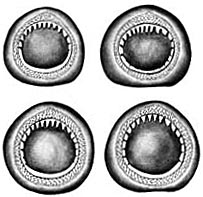
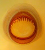
Figure. Arms and arm suckers of Mt. pyrodes. Left - Lateral and oral views of two arms, 65 mm ML, immature female, NMNH 729894. Photograph by R. Young. Middle - Oral views of large suckers of arms I-!V, respectively, relative sizes maintained, holotype. Drawings from Young (1972). Right - Oral view of large arm sucker, 102 mm ML, immature female, 28°53'N, 118°12'W, preserved, NMNH 727463.. Photograph by R. Young.
- Tentacles
- Tentacles whip-like, circular in cross-section.
- Tentacular clubs with microscopic protective membranes with tiny trabeculae.
- Tentacular club base abrupt (ie, suckers do not trail down stalk).
- Club suckers slightly elongate with 2-3 large lateral knobs projecting into orifice of sucker from outer ring; few tiny distal teeth on inner ring.
- Funnel
- Deep pocket between funnel bridles present. Click on an image to view larger version & data in a new window
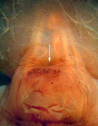
Figure. Ventral view of funnel and bridles showing deep pocket (arrow points to the opening of the pocket) between bridles, Mt. pyrodes, paratype, NMNH, 80 mm ML, 29°19'N, 119°21'W. Note photophores on dorsal surface of funnel. Photograph by R. Young.
- Fins
- Fins approximately circular, with small anterior lobes.
- Short tail present, usually broken.
- Photophores
- Integumental photophores present. Ringed chromatophores and white spheres absent. Compare with species of Mastigoteuthis here. Click on an image to view larger version & data in a new window
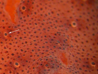
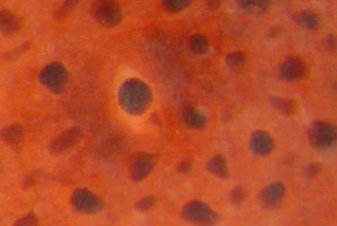
Figure. Ventral view of skin on the ventral surface of the head, Mt. pyrodes, 145 mm ML, nearly mature male, 31°53'N. 121°13'W. Left - Low magnification. Arrow points to photophore enlarged at the right. Right - High magnification of one photophore and surrounding tissue. Photograph by R. Young.
- Integumental photophores present on arms III and IV (at least) and on dorsal and ventral surfaces of head, funnel, mantle and fins; photophores more abundant on ventral surfaces; photophores present along entire ventral mantle extension between fins. Click on an image to view larger version & data in a new window
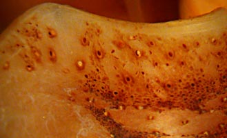
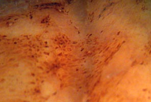
Figure. Integumental mantle photophores of Mt. pyrodes. Left - Ventral mantle margin showing photophores and chromatophores, NMNH 729894. Right - middorsal mantle (depression overiies gladius) near anterior end. In spite of damage, some dorsal photophores are visible. Photograph by R. Young.
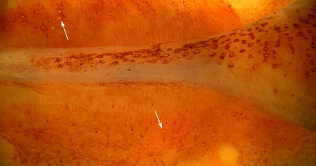
Figure. Ventral view of ventral fin photophores (arrows) of Mt. pyrodes, paratype, NMNH, 80 mm ML, 29°19'N, 119°21'W. Note also numerous photophores on the ventral mantle extension between the fins. Photograph by R. Young.
- Arms IV with integumental photophores in 3-5 irregular series. Click on an image to view larger version & data in a new window
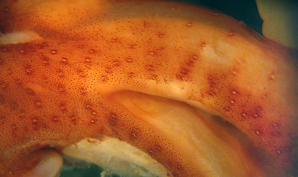
Figure. Ventral view of head and proximal arms IV, Mt. pyrodes, 145 mm ML, nearly mature male, 31°53'N. 121°13'W. Photograph by R. Young.
Click on an image to view larger version & data in a new window Click on an image to view larger version & data in a new window
Figure. Mt. pyrodes, 145 mm ML, same squid as above. Left - Twisted ventral view of arms IV, Arrow points to midline of the higher magnification picture at the right. Right - Higher magnification of a mid-region of arm IV. Note multiple series of arm IV photophores. Photographs by R. Young.
- Pigmentation
- Reddish dermal pigmentation overlaid by darker red chromatophores (see photographs above).
- Measurements
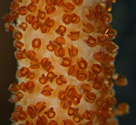
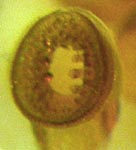
Figure. Tentacular club and club suckers of Mt. pyrodes, 102 mm ML, NMNH 727463. Left - Oral view of midregion of club. Right - Oral view of large club sucker. Photographs by R. Young.

Figure. Tentacular club and club suckers of Mt. pyrodes, holotype, 110 mm ML, male. A - Oral view of club sucker. B - Oral view of inner ring of club sucker. C - Oral view of club base. D - Aboral view of tentacular club. Drawings from Young (1972).
Scanning electron micrographs of the suckers can be seen here.
| Holotype | Paratype | Paratype | ||
| Sex | Male | Female | Male | Spent female |
| Mantle lenght | 110 | 148 | 86 | 145 |
| Mantle width | 27 | 34 | -- | |
| Fin length | 64 | 89 | 50 | 74 |
| Fin width | 61 | 90 | 50 | 70 |
| Arm I, length | 42 | 62 | 33 | |
| Arm II, length | 55 | 78 | 47 | |
| Arm III, length | 52 | 74 | 44 | |
| Arm IV, length | 99 | 120 | 79 | |
| Tentacle length | 233 | 378 | 188 | |
| Club length | 121 | 150 | 90 | |
| Eye diameter | 15 | 21 | 14 | |
| Tail length | 17+ | 20+ | 12+ | 18+ |
| Club sucker diameter | 0.28-0.32 | 0.30-0.35 | 0.24-0.26 |


