
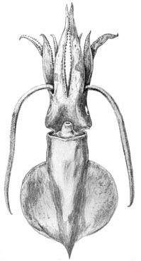
Figure. Ventral views of I. cordiformis. Left - Drawing from Sasaki (1929). Middle - Holotype, 80 mm ML. Right - Drawing from Voss (1963); dots on drawing represent skin tubercules.
- Arms
- Arm suckers with with teeth that "look like indentations" on distal margin of sucker-rings (Chun, 1910).
- Arm formula: 4>2>3>1. Suckers in two series.
- Arm I and III with ca 52 pairs of suckers; arm II with ca. 60 pairs and arm IV with ca. 64 pairs (Sasaki, 1929).
- Arm suckers:
- Tentacles
- Tentacular club not expanded (slightly expanded according to Sasaki, 1929, and Voss, 1963).
- Club with well-developed (for a mastigoteuthid) trabeculate protective membranes. Click on an image to view larger version & data in a new window
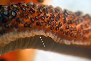
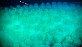
Figure. The club of I. cordiformis showing the well-developed trabeculate protective membrane (arrows). Left - Lateral view of a portion of a club, 70 mm ML, Japanese waters. Right - Oral view of a portion of a club, holotype, 80 mm ML. Photographs by R. Young.
- At club base suckers in two series (5 pairs of suckers), then three series, then increasing to 25 suckers across in middle of club (Chun, 1910).
- In the middle of the club, medial three series of suckers slightly larger than lateral ones (Chun, 1910)..
- Large club suckers 0.5 mm in diameter, holotype (Chun, 1910).
- Club sucker-rings toothed all around, with very long, distal teeth that extend orally rather than toward the center of the aperature (Chun, 1910).
- Head and funnel
- Funnel locking-apparatus "oval, with a groove"(Voss, 1963); "locking groove...rather deep, L-shaped, the tragus being well developed, while the antitragus is almost obliterated, being decidedly fainter than illustrated by Chun" (Sasaki, 1929).
- Nuchal cartilage spatulate (Chun, 1910).
- Funnel adductors do not project externally (Chun, 1910).
- Funnel adductors without medial pocket.
- Funnel locking-apparatus with well-developed tragus and much smaller antitragus. "The groove of the cartilage is divided posteriorly."
- Fins
- 70-80% of body length.
- Pigmentation
- Pigment appears to be mostly in chromatophores.
- Color according to ...
- Measurements and counts
Source Chun, 1910 Voss, 1963 Sasaki, 1929 Sex Male Male Gladius length 83 Mantle length 80 (ventral ML) 60 90 Mantle width 27 16 27 Tail 9 Fin Length 62 (with tail) 47 70 Fin Width 60 40 67 Head length Head width 28 20 28 Length, Arm I -- Left/Right 36 42 38/41 Length, Arm II 46 45 52/52 Length, Arm III 38 48 42/42 Length, Arm IV 59 70 60/60 Tentacle length 97 90/100 Club length 61 23* 60/60 Club sucker diameter 0.12 *Probable error: discrepancy between text and table in Voss (1963).

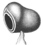
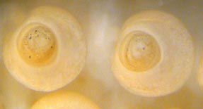
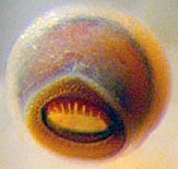
Figure. Arm suckers of I. cordiformis. Left - Oral and lateral views, holotype. Drawings from Chun (1910). Middle - Oral view, holotype. Right - Oral-distal view, 70 mm ML, Japanese waters. Photographs by R. Young.
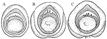
Figure. Oral views of arm suckers (Figs. A-C above) of I. cordiformis. Drawings from Voss (1963). Fig. A - Near arm base sucker rings nearly smooth with ca. 7 low, square teeth. Fig. B - Arm sucker of 7th row with single, slender median tooth and several blunt laterals. Fig. C - Arm sucker of 11 th row with 8-9 teeth, several being slender and square cut.
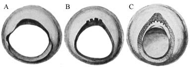
Figure. Oral views of arm suckers (Figs. A-C above) of I. cordiformis. Drawings from Sasaki (1929). Fig. A - Basal 3-4 arm sucker rows with smooth rings. Fig. B - Arm suckers in row 7 with 4-6 close set teeth on distal margin. Fig. C - Arm suckers in row 11 and distally with 8-11 teeth on distal margin.
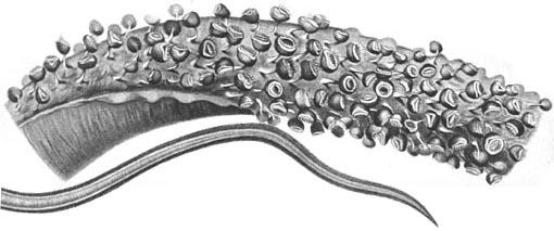
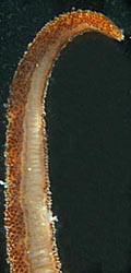
Figure. Tentacular club of I. cordiformis. Left top - Lateral to oral view of the club base, holotype. Left bottom - Aboral view of the tip of the club, holotype. Drawings from Chun (1910). Right - Aboral view of the club tip, 70 mm ML, Japanese waters. Note that the suckers do not completely encircle the club at the tip. Photograph by R. Young.
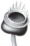
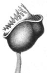
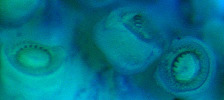
Figure. Oral and lateral views of club suckers of I. cordiformis, holotype. Left - Drawings from Chun (1910). Right - Photograph by R. Young. Suckers stained with methylene blue.
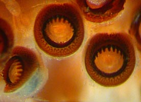
Figure. Oral view of club suckers, 70 mm ML, Japanese waters. Photograph by R. Young.
Voss (1963): Club slightly expanded; 38% of ML, 50% of tentacle length. Suckers in about 20 rows in club midpart. Club sucker ring with slender, square-tipped teeth all around; teeth long, slender distally, short proximally.Sasaki (1929): Club slightly expanded. Club length 2/3 to 3/5 of tentacle length. Suckers in ca. 8 series near base, shortly increasing to 12 series and reaching maximum of 24 from middle of club to tip. Large proximal club suckers with 20 or more blunt, separate teeth all around, distal teeth larger (but not as long and recurved as illustrated by Chun). Small distal suckers with 7-10 teeth on distal margin only.
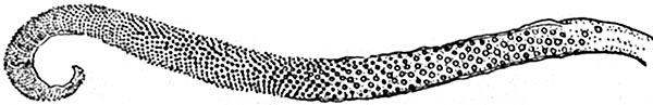
Figure. Oral view of tentacular club of I. cordiformis. Drawing from Voss (1963).
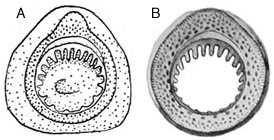
Figure. Oral view of club suckers of I. cordifomis. A - Drawing from Voss (1963). B - Drawing from Sasaki (1929).
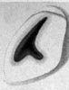
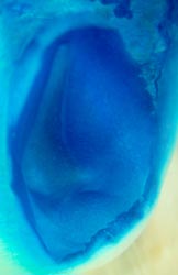
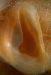
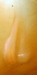
Figure. Funnel/mantle locking-apparatus of I. cordiformis. Left - Frontal view of funnel component, holotype. Drawing from Chun (1910). Middle - Frontal view of the funnel component, holotype. Stained with methylene blue. Photograph by R. Young. Right two photographs - Frontal views of the funnel and mantle components, 70 mm ML, Japanese waters. Note the unusual form of the mantle component. Photograph by R. Young.
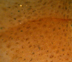
Figure. Skin of I. cordiformis, 70 mm ML, Japanese waters showing chromatophores. Photograph by R. Young.
- Chun (1910): Pale flesh-pink.
Voss (1963): Yellowish with small reddish-brown chromatophores.
Sasaki (1929): Thickly crowded deep brownish chromatophores.
Measurements in mm.


