
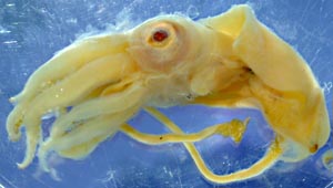
Figure. A. antarcticus, side view, 32 mm ML, preserved. Photograph by R. Young.
- Arms
- Arms II-IV subequal, arms I distinctly shorter.
- Arms IV more slender and less muscular than other arms; suckers much smaller than those of other arms (i.e., largest arms IV sucker about half the diameter of largest sucker of arm III).
- Most large suckers of arms I-III with single, large distal tooth, with smaller, less regularly shaped teeth to either side.
- Largest suckers of arms III with only distal tooth.
- Large distal tooth more prominent on arm suckers of larger specimens.
- Distal suckers with teeth of similar size along distal half of sucker ring.
- Tentacles
- Proximal manus
- eight of nine carpal suckers and knobs present along dorsal margin of proximal manus. Arrows in photograph on the right point to two carpal knobs (white arrows) and two carpal suckers (yellow arrows).
- Proximal to manus, carpal locking suckers and knobs cross to mid-oral side of tentacle stalk; broadly spaced suckers (with knobs) extend proximally most of tentacle length with suckers alternating on opposite sides of midline.
- Suckers of proximal manus become progressively larger towards distal end.
- Transversely arranged suckers of proximal manus number ca. 10-15; longitudinally arranged suckers number ca. 15-20.
- Proximal manus ca. 20-30% of club length, 25-40% of manus length.
- Distal manus
- Distal manus with two medial series of suckers enlarged to greatly enlarged. Each series with about 5 suckers.
- Marginal suckers about 1/3 diameter of largest medial suckers; marginal suckers about the same size as large suckers of dactylus.
- Trabeculate protective membranes present.
- Dactylus
- Club dactylus with three series of suckers; suckers progressively larger from dorsal to ventral series.
- Terminal pad with suckers present. Some terminal-pad suckers are missing in the photograph to the left.
- Dorsal keel present.
- Sucker dentition
- The large suckers of the distal manus have about 60 small, slender, pointed teeth around the entire margins.
- Similar but less numerous teeth are present on the suckers of the proximal manus and dactylus.
- Head
- Eyes large (diameter ca. 25% of ML).
- Olfactory organ a simple finger-like papilla.
- Radula, apparently homodont, with very slender teeth.
- Fins
- Anterior lobes absent; posterior lobes present.
- Length ca. 35% of ML.
- Gladius
- Gladius lacks conus; posterior edges of vanes fold ventrally to nearly touch.
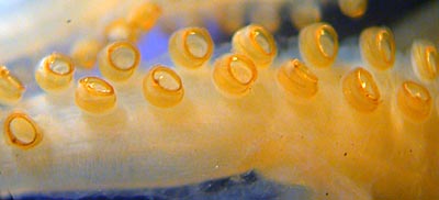
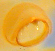
Figure. Left - Oral-lateral view of proximal portion of arm III of A. antarcticus, 32 mm ML. Right - Oral-distal view of large sucker from arm III of A. antarcticus, 32 mm ML.
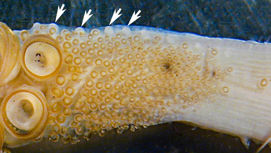
Figure. Oral view of proximal manus of A. antarcticus, preserved. Arrows point to some suckers (yellow) and knobs (white) of the proximal locking-apparatus of the club. Photograph by R. Young.
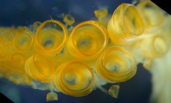
Figure. Oral view of distal manus of A. antarcticus, 32 mm ML, preserved. Photograph by R. Young.
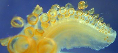
Figure. Oral view of dactylus of A. antarcticus, 32 mm ML, preserved. Photograph by R. Young.
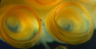

Figure. Left - Oral view of large suckers from the distal manus of A. antarcticus, 32 mm ML. Right - Oral view of suckers of the distal region of the proximal manus of A. antarcticus, 32 mm ML. Photographs by R. Young.

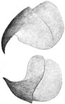
Figure. Left - Radula of A. antarcticus, 14.5 mm ML. Drawing modified from Odhner (1923). Right - Side view of beaks of A. antarcticus, upper beak (top) and lower beak (bottom). Drawing from Odhner (1923).
Figure. Posterior (left) and ventral views (right) of the gladius of A. antarcticus, 14.5 mm ML. Drawing from Odhner (1923).
Comments
Except for the photograph of the proximal manus and the drawings, the above description was taken from a 32 mm ML squid pictured in the title photograph. The peculiar posterior end of the gladius illustrated by Odhner (1923) was thought by Toll (1982) to have resulted from damage. Our examination indicates, however, that Odhner's illustration is accurate. Odhner had two specimens of nearly the same size (28 and 26 mm total length). We assume that all illustrations are from the larger specimen (14.5 mm GL=ML).


