- Arms
- Arms I 54-65% of ML.
- Arms III 73-103% of ML.
- Arms IV 120-200% of ML.
- Large arm suckers with 10-20 usually separate, pointed to blunt teeth on distal 2/3 of ring.
- Largest suckers not globular.
Scanning electron micrographs of the arm suckers can be seen here.
- Tentacles
- Club length 115 % of ML.
- Club suckers with 11-15 pointed teeth, in contact at bases, over distal 3/4 of ring; enlarged central tooth.
- Sucker stalks divided into two parts with flag-like lateral keel on proximal segment; stalks of suckers in lateral series about twice as long as those of medial series.
- Club protective membranes
- Membranes in two sets with proximal set only slightly broader than distal set.
- Proximal set short(ca. 7% of club length) with 11-13 trabeculae; at broadest point, two trabeculae occur between adjacent lateral suckers
- Distal set long (ca. 93% of club length) with 83-96 broad but separate ( < half the trabecula width), mostly undivided trabeculae (first trabecula may be divided).
Scanning electron micrographs of the club suckers can be seen here.
 Click on an image to view larger version & data in a new window
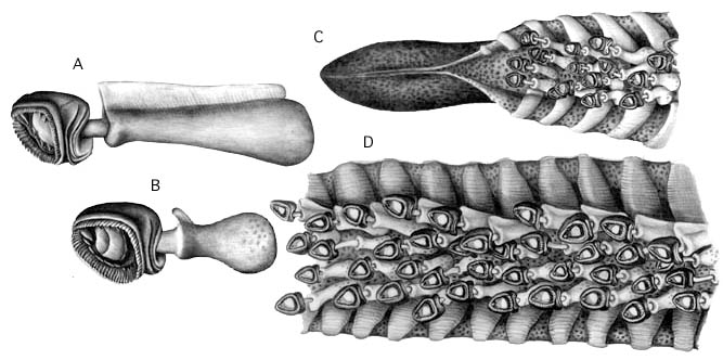
Figure. Oral views of portions of the tentacular club and club suckers of C. picteti. A - Lateral club sucker. Note the distinct "flag." B - Medial club sucker. Note the small size of stalk. C - Terminal portion showing club-tip photophore with lids closed. D - Midsection of the club showing the shape of trabeculae. Drawings by J. R. Schroeder.
Click on an image to view larger version & data in a new window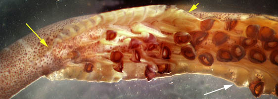
Figure. Photomicrograph of the base of the club of C. picteti, Hawaiian waters, preserved. The yellow arrows mark the approximate beginning and end of the proximal protective membrane. The white arrow points to a typical trabecula on the distal protective membrane. Photograph by R. Young.
Click on an image to view larger version & data in a new window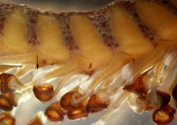
Figure. Lateral view of the distal portion of the club of C. picteti, Hawaiian waters. The arrow points to a flag on the sucker stalk. Photograph by R. Young.
- Head
- Head length 32-50% of ML.
- Head length 32-50% of ML.
- Funnel
- Funnel-locking apparatus: wide, low tragus that slightly undercuts mantle component; broad antitragus that deeply undercuts mantle component.
Click on an image to view larger version & data in a new window
Figure. Frontal view of the funnel locking-apparatus of C. picteti. Drawing by J. R. Schroeder.
- Fins
- Fin length ca. 55% of ML.
- Fin width ca. 45% of ML.
- Photophores
- Eyeball- 3 series of round organs: lateral series= 6 to 9; intermediate series = 8 to 11; medial series = 6 to 10; terminal photophores slightly larger.
- Viscera: two organs.
- Club-tip photophore small (exposed photogenetic surface much less than half of photophore length), with little or no terminal papilla.
- Aboral tentacular stalk with cushion-like photophores.
- Distal-most pad-like photophore partially embedded and located proximal to first club sucker.
Click on an image to view larger version & data in a new window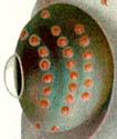
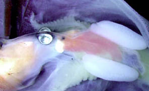
Figure. Photophores of C. picteti. Left - Ventral view of the ocular photophores. Drawing from Chun (1910). Right - Ventral view of the opened mantle cavity showing the visceral photophores on either side of the intestine, Hawaiian waters. Note also the large, white nidamental glands on this nearly mature female. Photograph by R. Young and C. Roper.
- Pigmentation
- Club-tip photophore with pigment in functional chromatophores.
- All pigment on clubs in chromatophore organs.
- Club sucker stalks without pigment.
- Buccal membrane usually with dense epidermal pigment.
- Olfactory organ with or without chromatophores depending on size.
Click on an image to view larger version & data in a new window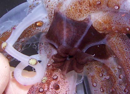
Figure. Oral view of the closed and heavily pigmented buccal membrane of a specimen of C. picteti from Hawaiian waters, central North Pacific. Note also the white tentaclular stalks with photophores, two of which are smaller than the others and have violet pigment.
Comments
The very short, proximal segment of the club protective membrane can be difficult to recognize as distinct from the distal segment.
The characters described above are based on several specimens from across the geographical range of C. picteti (off New Zealand, Hawaii, Japan and Indonesia) and the holotype of C. imperator (= synonym) from off Sumatra, Indonesia.
We found considerable geographical variation in the following characters: size of the tentacular club; number of trabeculae proximal to the first club sucker; club sucker dentition; arm-sucker dentition; position of the distal stalk photophores; number of arm suckers; number of ventral arm photophores; sucker size; buccal membrane pigmentation.


