Stoloteuthis maoria
Richard E. Young, Michael Vecchione, and Clyde F. E. RoperIntroduction
Stoloteuthis maoria was previously placed in Iridoteuthis but we have transferred it to Stoloteuthis due to the similarity in its armature to S. leucoptera. Very little is known about its biology.
Brief diagnosis:
A Stoloteuthis with ...
- broad head-mantle fusion.
- glandular structures only on ventral borders of arms I in males.
Characteristics
- Arms
- Arms III with large, gelatinous aboral keels; arms IV with lateral keels.
- Arm formula: II>=III>I>=IV.
- Arms with delicate, trabeculate protective membranes.
- Arms I, II and III joined by extensive web, greatest in sector A, smallest in sector C.
- Arms with two series of globular suckers.
- In males, with few suckers in mid-arm II greatly enlarged; both arms I with expanded protective membrane on proximal 2/3 of ventral border which contain a series of glands with small pores at their medial ends.
- Arm suckers with smooth inner rings.
- Tentacles
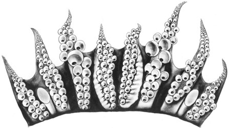
- Tentacular clubs short, unexpanded, with about 18-22 suckers in a transverse series; suckers at club base enlarged, often about 16-18 in number (range at least 13-27). Suckers adjacent to proximal patch of suckers, very small, become progressively larger distally with largest in distal half of club.
- Protective membranes and aboral keels absent.
- Tentacular organ present.
- Oral face of tentacular stalk flattened.
 Click on an image to view larger version & data in a new window
Figure. Oral view of the tentacular club of S. maoria, mature male, 14 mm ML, off S.E. New Zealand. Drawing by A. Hart.
- Tentacular clubs short, unexpanded, with about 18-22 suckers in a transverse series; suckers at club base enlarged, often about 16-18 in number (range at least 13-27). Suckers adjacent to proximal patch of suckers, very small, become progressively larger distally with largest in distal half of club.
- Head
- Eyes large, with cornea and secondary eyelids.
- Olfactory organ a short, thick cyclinder with terminal concavity.
- Funnel
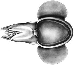
- Funnel large, nearly covered by the ventral mantle.
- Funnel component of the funnel/mantle locking apparatus with straight groove; mantle component with straight ridge termininating in a slight depression.
- Lateral funnel adductor muscles present.
- Dorsal pad of funnel organ with inverted V-shape and small anterior papilla; ventral pads oval, very large, cover entire internal ventral surface of funnel.
- Funnel valve present.
- Mantle
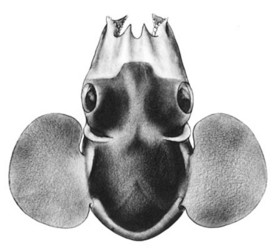
- Mantle broadly fused to head dorsally to a level in line with the dorsal edges of the eye lenses.
- Mantle excavated on its midventral margin.
- Ventral shield extends for about 90% of the ventral mantle length.
 Click on an image to view larger version & data in a new window
Figure. Ventral view of S. maoria, mature male, 14 mm ML, off S.E. New Zealand. Drawing by A. Hart.
- Fins
- Fins large, about equal to the mantle length.
- Fins extend anteriorly beyond the lateral fusion of the mantle.
- According to Harman and Seki (1990), fins have pointed posterior lobes.
- Pigmentation
- A silver band passes around lateral and posterior surfaces of mantle, above and below the fins except for a small area dorsal to the fins. A silver area from the anterior edge of each eye extends to the base of arms II and dorsally over the web to cover about half of the web in the dorsal midline.
- A silver band passes around lateral and posterior surfaces of mantle, above and below the fins except for a small area dorsal to the fins. A silver area from the anterior edge of each eye extends to the base of arms II and dorsally over the web to cover about half of the web in the dorsal midline.
- Gladius
- Gladius absent.
- Gladius absent.
- Viscera
- Mature eggs about 2 mm in longest diameter.
- Mature eggs about 2 mm in longest diameter.
- Measurements: Sex - Male, DML - 14, Fin L - 14, Fin W - 9, Arm I L - 8, Arm II L - 9, Arm III L - 9, Arm IV L - 7.5, Web A - 6, Web B - 5, Web C - 4.
Comments
The illustrations were made many years ago, and the description is taken from even older notes. We have not compared S. leucoptera and S. maoria side by side. Differences between the two species need to be confirmed.
S. maoria differs from S. leucoptera in the broader fusion of the head and mantle, glands only on the ventral sides of arms I in males rather than both sides, arms IV tips with two series of suckers rather than 3-4 and the apparently pointed posterior fin lobes. S. maoria differs from S. weberi in having the mantle and head fused dorsally rather than having the mantle margin free.
Life History
In all four mature females examined spermatangia were found lodged in the gelatinous tissue covering the arms, head and neck regions on only the left side of each animal.
Distribution
Type locality - Paraparaumu Beach (washed ashore), Wellington, New Zealand. Paratypes from 41°31'S, 174°58'W and Cook Straight, New Zealand from 240-370 m.
Our specimens came from 44° 02'S, 178°08'W (Chatham Rise off New Zealand) and 51°12'S, 166°31'E (off the southwestern tip of New Zealand) over bottom depths of 230-573 m.
References
Harman, R. F. and M. P. Seki. 1990. Iridoteuthis iris (Cephalopoda: Sepiolidae): New records from the central North Pacific and first description of the adults. Pac. Sci. 44: 171-179.
About This Page

University of Hawaii, Honolulu, HI, USA

National Museum of Natural History, Washington, D. C. , USA

Smithsonian Institution, Washington, D. C., USA
Page copyright © 2014 , , and
 Page: Tree of Life
Stoloteuthis maoria .
Authored by
Richard E. Young, Michael Vecchione, and Clyde F. E. Roper.
The TEXT of this page is licensed under the
Creative Commons Attribution-NonCommercial License - Version 3.0. Note that images and other media
featured on this page are each governed by their own license, and they may or may not be available
for reuse. Click on an image or a media link to access the media data window, which provides the
relevant licensing information. For the general terms and conditions of ToL material reuse and
redistribution, please see the Tree of Life Copyright
Policies.
- First online 26 December 2007
- Content changed 26 December 2007
Citing this page:
Young, Richard E., Michael Vecchione, and Clyde F. E. Roper. 2007. Stoloteuthis maoria . Version 26 December 2007 (under construction). http://tolweb.org/Stoloteuthis_maoria/27775/2007.12.26 in The Tree of Life Web Project, http://tolweb.org/


 Go to quick links
Go to quick search
Go to navigation for this section of the ToL site
Go to detailed links for the ToL site