


Eucleoteuthis
Eucleoteuthis luminosa
Richard E. Young and Michael Vecchione

Introduction
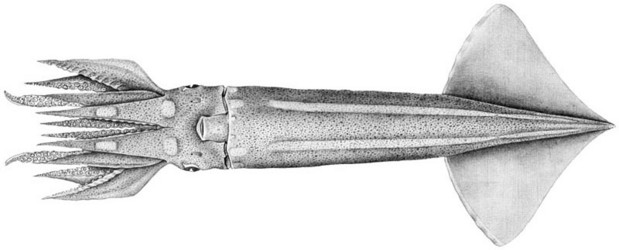
Eucleoteuthis luminosa is a very distinctive squid with two long luminous stripes on the ventral mantle surface and fused funnel/mantle locking-apparatus. The squid is intermediate in size, rarely exceeding 170 mm ML (Wormuth, 1976).
Brief diagnosis:
An ommastrephid ...
- with 2 longitudinal strips of luminous tissue on ventral mantle extending nearly entire length of mantle.
Characteristics
- Arms
- Left arm IV hectocotylized:
- Proximal two thirds with about 19-22 suckers in two series; protective membranes weak.
- Distal tip of hectocotylus with two series of pointed knobs in place of suckers.
- Distal tip of hectocotylus with ventral protective membrand more developed than dorsal membrane. (Wormuth, 1975)
Click on an image to view larger version & data in a new window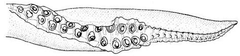
Figure. Oral view of the hectocotylus of E. luminosa. Drawing from Roeleveld (1988).
- Left arm IV hectocotylized:
- Tentacles
- Largest club suckers with smooth or slightly dentate ring with 1 large tooth.
Click on an image to view larger version & data in a new windowClick on an image to view larger version & data in a new window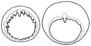
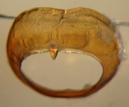
Figure. Oral view of club-manus sucker rings of E. luminosa. Left - More proximal ring. Middle - Largest ring. Both drawings from Roeleveld (1988). Right - Tilted oral view of the largest ring. Photograph by R. Young
- Largest club suckers with smooth or slightly dentate ring with 1 large tooth.
- Head
- Beaks: Descriptions can be found here: Lower beak; upper beak.
- Beaks: Descriptions can be found here: Lower beak; upper beak.
- Funnel
- Mantle locking-apparatus with anterior bifurcation.
- Mantle and funnel components of locking-apparatus fused proximally.
Click on an image to view larger version & data in a new window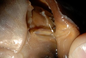
Figure. Anterior view of the funnel/mantle locking apparatus of E. luminosa with the mantle folded back to show the region of fusion (arrow). Photograph by R. Young.
Click on an image to view larger version & data in a new window
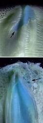
Figure. Funnel/mantle locking apparatus of E. luminosa. Left - Funnel component. Middle - Mantle component. Note cut portion of lock where fusion occurred in both photographs. Upper right - Anterior tip of mantle component. Arrow points to cartilage betweem limbs of the bifurcation. Lower right - Anterior tip of mantle component. Arrow points to appendix. Photographs by R. Young.
- Mantle locking-apparatus with anterior bifurcation.
- Photophores
- Small subcutaneous photophores on ventral surfaces of mantle, head and arms III-IV.
- Ventral surface of mantle with 2 longitudinal strips of luminous tissue extending nearly entire length of mantle.
- Two large, oval luminous patches anterior to each mantle strip.
- Arm IV with one large oval luminous patch at arm base and another midway to arm tip.
Click on an image to view larger version & data in a new window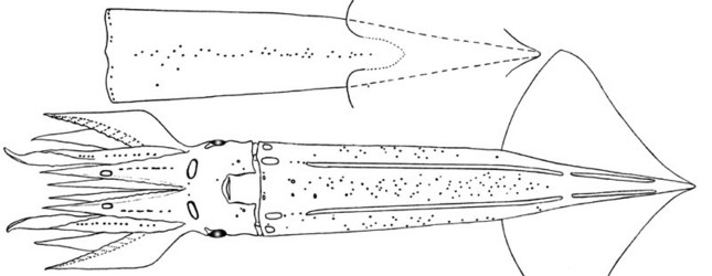
Figure. Photophore arrangement of E. luminosa. Top - Photophores of the dorsal mantle. Bottom - Ventral photophores. Drawings from Young (1972).
Click on an image to view larger version & data in a new windowClick on an image to view larger version & data in a new window
Figure. Ventral view of the head and anterior mantle of E. luminosa showing large photophore patches. Two small patches are present near the anterior mantle margin on either side of the midline. The white lines are from surface reflection as the photograph was taken with the squid out of water. Photograph by M. Vecchione.
Comments
More details of the description of E. luminosa can be found here.
Life History
Toshe Wakabayashi and Richard E. Young
The following description is from Wakabayashi et al. (2002).
- Proboscis
- Proboscis long; PI (Proboscis Index - ratio between proboscis length and mantle length) = 0.77 at 2.2 mm ML and 0.73 at 4.4 mm ML.
- Lateral proboscis suckers 1.3 - 1.9 times (mean = 1.6) diameter of medial suckers. Click on an image to view larger version & data in a new window
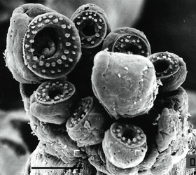
Figure. Distal view of the tip of the proboscis of an 5.5 mm ML paralarva of E. luminosa showing enlarged lateral suckers. Scale bar = 100 µm. Scanning electron micrograph from Wakabayashi et al. (2002).
- Proboscis split to form two tentacles at 8.6 mm ML. The proboscis grows rapidly to about 6 mm ML then begins to shrink until the tentacles form from the spliting of the proboscis along its midline. The tentacles then grow rapidly. Click on an image to view larger version & data in a new window

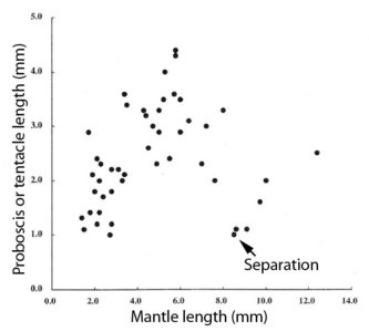
Figure. Left - Oral views of the brachial crown showing the proboscis starting to split apart at its base (left, 6.4 mm ML) and the split just completed to form tentacles (right, 9.1 mm ML). Bar = 1 mm. Note the extremely small size of the newly formed tentacles. Right - Diagram showing the growth of the proboscis against mantle length. Figures and diagram from Wakabayashi et al. (2002).
- Photophores
- Single intestinal photophore present at 3.3 mm ML.
- Single, round photophore present on ventral surface of each eye at 4.4 mm ML.
- Funnel
- Funnel/mantle locking-apparatus not fused until 9-10 mm ML.
- Funnel/mantle locking-apparatus not fused until 9-10 mm ML.
- Chromatophores
- At 2.2 mm ML (see illustration above):
- Dorsal mantle - 4 chromatophores (C) in irregular, transverse row in mid-mantle; single C at posterior end.
- Ventral mantle - 4 C in trapezoid pattern.
- Head - 1 or 2 pairs C dorsally; 1 pair on posteroventral margin.
- At 4.4 mm ML (see illustration above):
- Dorsal mantle - Same except 5 C.
- Ventral mantle - Same but with 5 C along anteroventral mantle margin and 1 pair at posterior end of mantle.
- At 5.0 mm ML:
- Head - 7-11 C dorsally, 4 ventrally.
- Arms - Arms I, II and III with 2, 5, 5 C respectively on aboral side.
- Buccal region - 1 pair C on outer lip by arms II and III.
- At 6.4 mm ML:
- Arms - Same.
- Buccal region - Same.
- At 9.1 mm ML:
- Buccal region - Same but with 1 C between arms I.
- Buccal region - Same but with 1 C between arms I.
- At 2.2 mm ML (see illustration above):

Figure. Oral (top) and dorsal/ventral (bottom) views of paralarvae of E. luminosa Note the ocular photophores and the chromatophore patterns. Scale bars = 1.0 mm. Drawings from Wakabayashi et al. (2002).
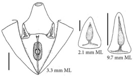
Figure. Paralarvae of E. luminosa. Left - Ventral view of the mantle cavity showing the intestinal photophore and funnel locking-apparatus. Right - Funnel locking-apparatus in paralarvae of two sizes. Scale bars = 1 mm. Drawings from Wakabayashi et al. (2002).
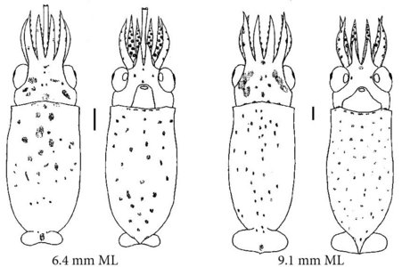
Figure. Oral (top) and dorsal/ventral (bottom) views of paralarvae of E. luminosa. Scale bars = 1.0 mm. Drawings from Wakabayashi et al. (2002).
Distribution
Type locality: Sagami Bay, Japan.
Subtropical to temperate waters of the Pacific, South Atlantic and Indian oceans: North Pacific from the northwest (25°-45°N) to northeast (15°-40°N); South Atlantic (10°-35°S); South Pacific and Indian Oceans (15-35°S) (Nesis, 1982/87).
References
Roeleveld, M. A. 1988. Generic interrelationships within the Ommastrephidae (Cephalopoda). P.277-314. In: M. R. Clarke and E. R. Trueman (eds.). The Mollusca. Vol. 12. Paleontology and Neontology of Cephalopods. Academic Press, N.Y., 355pp.
Wakabayashi, T., K. Saito, K. Tsuchiya and S. Segawa. 2002. Descriptions of Eucleoteuthis luminosa (Sasaki, 1915) and Ornithoteuthis volatilis (Sasaki, 1915) paralarvae in the Northwestern Pacific. Venus, 60 (4): 237-260.
Young, R. E. 1972. The systematics and areal distribution of pelagic cephalopods from the seas off Southern California. Smithson. Contr. Zool., 97: 1-159.
Title Illustrations
| Scientific Name | Eucleoteuthis luminosa |
|---|---|
| Location | Hawaiian waters |
| Specimen Condition | Fresh |
| View | Ventral |
| Size | 187 mm ML |
| Image Use |
 This media file is licensed under the Creative Commons Attribution-NonCommercial License - Version 3.0. This media file is licensed under the Creative Commons Attribution-NonCommercial License - Version 3.0.
|
| Copyright |
©

|

| Scientific Name | Eucleoteuthis luminosa |
|---|---|
| Location | Off Southern California |
| Reference | Young, R. E. 1972. The systematics and areal distribution of pelagic cephalopods from the seas off Southern California. Smithson. Contr. Zool., 97: 1-159. |
| View | Ventral |
| Size | 163 mm ML |
| Image Use |
This media file is licensed under the Creative Commons Attribution-NonCommercial License - Version 3.0.
|
| Copyright |
©
|
About This Page
University of Hawaii, Honolulu, HI, USA
National Museum of Natural History, Washington, D. C. , USA
Page copyright © 2018 and
 Page: Tree of Life
Eucleoteuthis . Eucleoteuthis luminosa .
Authored by
Richard E. Young and Michael Vecchione.
The TEXT of this page is licensed under the
Creative Commons Attribution-NonCommercial License - Version 3.0. Note that images and other media
featured on this page are each governed by their own license, and they may or may not be available
for reuse. Click on an image or a media link to access the media data window, which provides the
relevant licensing information. For the general terms and conditions of ToL material reuse and
redistribution, please see the Tree of Life Copyright
Policies.
Page: Tree of Life
Eucleoteuthis . Eucleoteuthis luminosa .
Authored by
Richard E. Young and Michael Vecchione.
The TEXT of this page is licensed under the
Creative Commons Attribution-NonCommercial License - Version 3.0. Note that images and other media
featured on this page are each governed by their own license, and they may or may not be available
for reuse. Click on an image or a media link to access the media data window, which provides the
relevant licensing information. For the general terms and conditions of ToL material reuse and
redistribution, please see the Tree of Life Copyright
Policies.
- First online 29 November 2009
- Content changed 31 October 2018
Citing this page:
Young, Richard E. and Michael Vecchione. 2018. Eucleoteuthis . Eucleoteuthis luminosa . Version 31 October 2018 (under construction). http://tolweb.org/Eucleoteuthis_luminosa/19949/2018.10.31 in The Tree of Life Web Project, http://tolweb.org/


