


Abralia heminuchalis
Kotaro Tsuchiya and Richard E. Young

Introduction
A. heminuchalis is a small sized species, less than 40mm DML, found in the tropical Pacific. It is very similar to A. siedleckyi from off South Africa and may prove to be a junior synonym of the latter. It is characterized by its proportionally short, robust mantle and a photophore arrangement on the ventral head and mantle in which large photophores have a space surrounding each organ.
Brief diagnosis:
An Abralia (Heterabralia) with ...
- anterior ocular photophore comparable in size to largest of 3 medial ocular photophores but much smaller than posterior ocular photophore.
- irregular photophore series on mid-region of arms IV.
Characteristics
- Tentacle clubs
- Two or three hooks on ventral side.
- Two rows of large suckers on dorsal side of manus.
Click on an image to view larger version & data in a new window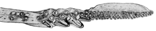
Figure. Oral view of the tentacular club of A. heminuchalis. Drawing from Burgess (1991, p. 121, Fig. 2J)
- Hectocotylus
- Left ventral arm of male hectocotylized.
- Hectocotylus with two different sized off-set flaps: an elongate, proximal flap on ventral margin and much shorter, distal flap on dorsal margin.
Click on an image to view larger version & data in a new window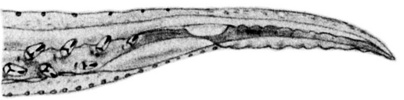
Figure. Oral view of the distal portion of the hectocotylus of A. heminuchalis. Drawing from Burgess (1991, p. 121, Fig. 2I).
- Eye Photophores
- Five, complex organs: two terminal opaque organs and three intermediate silvery organs. Posterior opaque organ much larger than others; anterior opaque photophore about the same size as the largest intermediate organ.Click on an image to view larger version & data in a new window

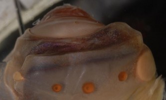
Figure. Ventral view of the ocular photophores of A. heminuchalis, anterior is at left. Top - Drawing from Burgess (1991, p. 121, Fig. 2B). Bottom - Photograph of a damaged, preserved eye. Note the pale color of the two terminal photophores and the orange color of the three medial photophores. Photograph by R. Young.
- Five, complex organs: two terminal opaque organs and three intermediate silvery organs. Posterior opaque organ much larger than others; anterior opaque photophore about the same size as the largest intermediate organ.
- Integumental Photophores
- Arms IV with scattered photophores between the two photophore series (the series on the edge of the lateral memberane constitutes a third series).
- Ventral mantle and head with scattered arrangement of integumental organs.
- Large organs each surrounded by space lacking photophores (black arrows in photograph). Their appearance (which is poorly defined in the photograph) varies depending on the contraction state of nearby chromatophores.
- Scattered, small photophores on midregion of arms IV between medial and lateral arm photophore series (white arrow in photograph). These photophores apprear at about 25 mm ML and are absent in A. andamanica and A. trigonura.
Click on an image to view larger version & data in a new window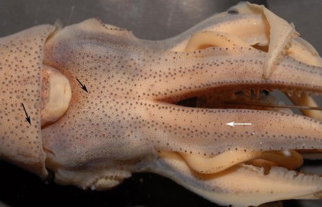
Figure. Ventral view of the head, posterior region of arms IV and anterior region of mantle of A. heminuchalis, 35 mm ML, equatorial pacific, mature male. Note the small photophores present in the mid-region of arms IV (white arrow). Note, also, the large photophores surrounded by a ring of smaller photophores, two of which are indicted by the black arrows. Photograph by R. Young.
- Reproductive structures
- Posterior region of nuchal cartilage replaced by part of the receptacle for spermatangia in females.
Click on an image to view larger version & data in a new window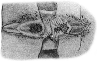
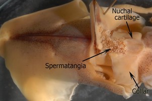
Figure. Dorsal (top) and dorsolateral (bottom) views of the receptacle for spermatangia in female A. heminuchalis. Anterior is right. Note that the posterior region of the nuchal cartilage is covered by the organ and that permatangia attach to ruguose areas (not a deep pit) on posterior portion of nuchal cartilage and gladius. Mantle, gladius and attachment area folded posteriorly. Drawing from Burgess (1991, p. 121, Fig. 2K). Photograph by R. Young.
- Posterior region of nuchal cartilage replaced by part of the receptacle for spermatangia in females.
Distribution
Geographical distribution
Distribution is the tropical Pacific from 5°N, 98°W (Okutani, 1974) to waters near Palau Isl. (6°N, 135°E) (Hidaka and Kubodera, 2000) and 16°S (Burgess, 1991) to 12°N (Hidaka and Kubodera, 2000).References
Burgess, L.A. 1992. Squids of the genus Abralia (Cephalopoda) from the central equatorial Pacific with a description of Abralia heminuchalis, new species. Bulletin of Marine Science, 49(1-2):113-136.
Hidaka, K. and T. Kubodera. 2000. Squids of the genus Abralia (Cephalopoda: Enoploteuthidae) from the western tropical Pacific with a description of Abralia omiae, a new species. Bulletin of Marine Science, 66(2):417-443.
Okutani, T. 1974. Epipelagic decapod cephalopods collected by micronekton tows during the EASTROPAC expeditions, 1967-1968 (systematic part). Bull. Tokai Reg. Fish. Res. Lab., 80: 29-118.
Title Illustrations
| Scientific Name | Abralia heminuchalis |
|---|---|
| Location | Equatorial Pacific |
| Comments | Drawing from Burgess (1991, p. 121, Fig. 2A) printed with the Permission of the Bulletin of Marine Science. |
| Reference | Burgess, L.A. 1992. Squids of the genus Abralia (Cephalopoda) from the central equatorial Pacific with a description of Abralia heminuchalis, new species. Bulletin of Marine Science, 49(1-2):113-136. |
| Sex | Female |
| View | Ventral |
| Size | 37 mm ML |
| Collection | USNM 729683 |
| Type | Holotype |
| Copyright | © Bulletin of Marine Science |
About This Page
Drawings from Burgess (1991) printed with the Permission of the Bulletin of Marine Science.

Tokyo University of Fisheries, Tokyo, Japan
University of Hawaii, Honolulu, HI, USA
Page copyright © 2018 and
 Page: Tree of Life
Abralia heminuchalis .
Authored by
Kotaro Tsuchiya and Richard E. Young.
The TEXT of this page is licensed under the
Creative Commons Attribution-NonCommercial License - Version 3.0. Note that images and other media
featured on this page are each governed by their own license, and they may or may not be available
for reuse. Click on an image or a media link to access the media data window, which provides the
relevant licensing information. For the general terms and conditions of ToL material reuse and
redistribution, please see the Tree of Life Copyright
Policies.
Page: Tree of Life
Abralia heminuchalis .
Authored by
Kotaro Tsuchiya and Richard E. Young.
The TEXT of this page is licensed under the
Creative Commons Attribution-NonCommercial License - Version 3.0. Note that images and other media
featured on this page are each governed by their own license, and they may or may not be available
for reuse. Click on an image or a media link to access the media data window, which provides the
relevant licensing information. For the general terms and conditions of ToL material reuse and
redistribution, please see the Tree of Life Copyright
Policies.
- Content changed 29 December 2009
Citing this page:
Tsuchiya, Kotaro and Richard E. Young. 2009. Abralia heminuchalis . Version 29 December 2009 (under construction). http://tolweb.org/Abralia_heminuchalis/19665/2009.12.29 in The Tree of Life Web Project, http://tolweb.org/


