


Stauroteuthidae
Stauroteuthis
Martin Collins, Richard E. Young, and Michael Vecchione
A single genus and two species are currently recognized in the family. However similar octopods from outside the Atlantic Ocean have not been critically examined.
- Stauroteuthis gilchristi (Robson, 1924)
- Stauroteuthis syrtensis Verrill, 1879
Introduction
Stauroteuthids are peculiar, gelatinous cirrates with a mantle opening that forms a complete tube around the funnel. They also have peculiar gills and internal shells and a large web that is nearly equally developed between all arms. When observed from submersibles, this octopod commonly has its arms and web formed into a bell-shape (bell-shape posture). Sometimes when the octopod is disturbed, it will inflate the web and draw the arms together at their tips to form a "balloon" with the arms and web (balloon posture). These postures are thought to be involved in feeding and/or defense (Vecchione and Young, 1997).
Brief diagnosis:
A cirrate with ...
- long cirri and secondary webs.
- U-shaped shell.
- tubular anterior mantle.
Characteristics
- External morphology
- Shape
- Anterior-posterior elongation of body pronounced; body extends well posterior to shell.
- Mantle
- Anterior mantle opening modified into a complete cyclinder formed by muscular mantle and encircling funnel base.(See "Comments" below.)
- Web
- Primary and secondary webs present.
- Web nodules absent.
- Cirri
- Cirri begin between suckers 2-6.
- Cirri length: Longest more than twice arm diameter.
- Cirri absent from arm tips. Cirri end before sucker 18-24.
- Suckers
- Suckers small, cylindrical.
- Suckers enlarged in male S. syrtensis.
- Beaks: See S. syrtensis.
- Shape
- Mantle cavity
- Mantle septum
- Thick, open posteriorly.
- Olfactory organ
- Lies well inside of mantle cavity from free mantle margin. (See "Comments" below.)
- Gills
- Gills have a diagnostic form. The secondary and tertiary lamellae are highly branching; the primary lamellae are difficult to recognize and don't form a symmetrical series along the gill; a large afferent vessel does not dominate the "top" of the gill. Click on an image to view larger version & data in a new window
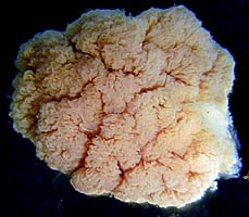
Figure. "Top" view (i.e. opposite the attachment to the mantle wall) of the preserved gill of S. syrtensis captured by submersible off Cape Hatteras, western North Atlantic. Photograph by R. Young.
- Gills have a diagnostic form. The secondary and tertiary lamellae are highly branching; the primary lamellae are difficult to recognize and don't form a symmetrical series along the gill; a large afferent vessel does not dominate the "top" of the gill.
- Internal anatomy
- Salivary glands
- "Anterior" salivary glands present.
- Radula
- Absent.
- Digestive gland
- Unilobular.
- Digestive tract
- Simple U-shaped digestive tract.
- Large posterior lip glands present. Click on an image to view larger version & data in a new window
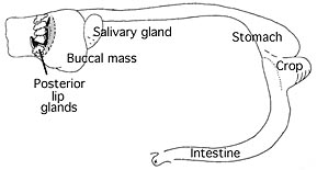
Figure. Lateral view of the digestive tract of S. syrtensis. Drawing modified from Vecchione and Young, 1997.
- Optic lobe
- Optic lobe spherical.
- Single optic nerve bundle passes through the white body. Click on an image to view larger version & data in a new window
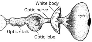
Figure. Dorsal views of the eye, optic lobe, white body and optic nerve of S. syrtensis. Drawing from Collins and Henriques, 2000.
- Salivary glands
- Mantle septum
- Shell
- The shell is U-shaped. Click on an image to view larger version & data in a new window
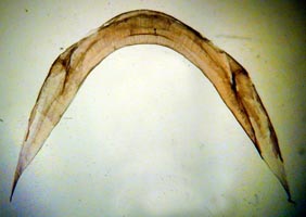
Figure. Anterior (?) view of the shell of a young S. syrtensis. Photograph by R. Young.
- The shell is U-shaped.
- Pigmentation
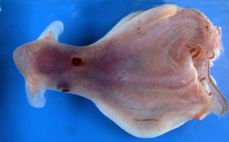
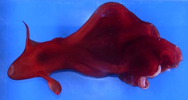
Figure. Views of S. syrtensis. Left - ventral view. Right - dorsal view. These two photographs show the color variation observed in two different fresh/living octopods of approximately the same size. The significance of this variation is unknown. Photographs by R. Young taken aboard the R/V G.O.SARS during the MARECO cruise to the central North Atlantic.
Comments
One of the most unusual features of the family is the cyclindrical, muscular mantle opening. Stauroteuthis spp. are the only cirrates in which the free edges of the mantle fuse dorsally to form a tube. The two photos taken at the right in a aquarium show the tubular opening in one stage of contraction. The opening appears like an elevated ring around the base of the slender, protruding funnel. Judging from preserved octopods (see drawing below), this ring can be greatly extended. In other octopods, the mantle attaches anteriorly, on either side, to the cephalic cartilage; as a result the dorsal wall of the mantle cavity, in this region, consists of the head.
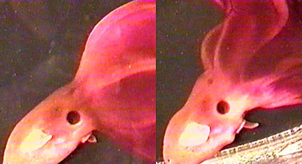
Figure. Lateral views of the head and mantle of S. syrtensis, alive. Video frames from a video taken by Edith Widder of the octopod in a planktonkreissel.
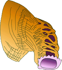
Figure. Twisting dorsolateral view of the anterior mantle of S. syrtensis showing the intertwining muscles that form the dorsal wall of the mantle at its anterior end. In this preserved octopod, the anterior mantle was contracted into a tube that, at first, appeared to be the funnel. The true funnel was withdrawn inside of the mantle cavity. The mantle opening, in this drawing, is colored purple and is at the lower right of the drawing. The skin has been removed showing the mantle muscles. Beneath the dorsal mantle muscles, the dark violet lining of the mantle cavity is seen.
In a fresh, translucent Stauroteuthis (below), the pigmented mantle cavity has the shape of a bow tie due to the large, central unpigmented region occupied by the thick mantle septum and the pigmented extensions of the mantle cavity anterolaterally leading to the dorsal mantle cavity.
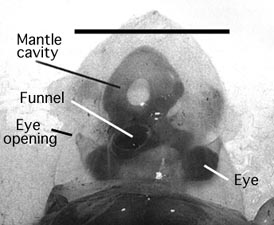
Figure. Mantle cavity of S. gilchristi, photograph modified from Collins and Henriques, 2000. Scale bar = 100 mm.
Comparisons Between Species
The two species are separated geographically (see below) but are very similar morphologically. Separation relies primarily on the differences in size and position of the suckers as seen in the following table:
| S. syrtensis Males |
S. syrtensis Females |
S. gilchristi Males |
S. gilchristi Females |
|
| Max. sucker diam., mm | 2.2-6 | 1.2-2.2 | 5.7-9.0 | 3.9-4.8 |
| Position of largest suckers | 12-16 | 1-3 | 9-14 | 9-14 |
| Sucker index (rel. to head W.) | 0.06-0.10 | 0.02-0.05 | 0.09-0.13 | 0.09-0.12 |
Nomenclature
Chunioteuthis Grimpe, 1916 is placed as a junior synonym of Stauroteuthis by Collins and Henriques (2000).
Behavior
Submersible observations have been made on S. syrtensis and we assume that they apply to both species. S. syrtensis is commonly seen, when first approached, with the web inflated into a bell-shape (below left) with the arms separated from the primary web by the secondary web. A ballooning posture is occasionally seen (below right), usually following disturbance, in which the web is extremely inflated and closed at the arm tips.
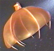
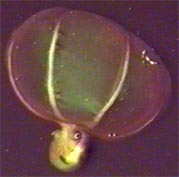
Figure. Lateral views of S. syrtensis. Left - Bell posture. Right - balloon posture. Submersible photographs.
Locomotion is via movement of the fins or the expulsion of water from the web. The latter method, however, is weak perhaps because of the loss of the strong connection of the web to the arms due to the presence of the secondary web. Small copepods have been found in the stomachs. This and the presence of large secretory lip glands suggest that mucous may be involved in the capture of prey. The above is summarized from Vecchione and Young, 1997.
Johnsen, et al. (1999) found that mechanical stimulation induced luminescence associated with the basal suckers of S. syrtensis (sex unknown). This is the only known case of bioluminescence in cirrate octopods and suggests that careful observations of living material may be useful. The function of the luminescence is unknown.
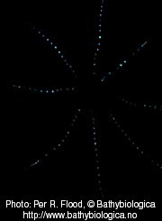
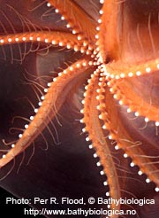
Figure. Left - Oral view of bioluminescence of S. syrtensis. Right - Oblique-oral view to show arrangement of suckers for comparison with Left photograph. Photographs by P. Flood who writes, "The bioluminescence was stimulated by brief electrical AC pulses from a 12V power supply unit. The bioluminescent suckers lit up one after the other in perfect succession from mouth to periphery when stimulated near the mouth area. They were never seen to flicker independent of their neighbours in the same row.
Distribution
Presently stauroteuthids are known only from the Atlantic Ocean. They are thought to be benthopelagic in distribution (Collins and Henriques, 2000). That is, they are pelagic but reside in close proximity to the ocean floor.
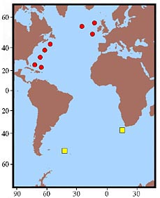
Figure. Distribution map, modified from Collins and Henriques, 2000. The red dots indicate capture locations of S. syrtensis and the yellow squares represent the capture locations of S. gilchristi.
References
Collins, M. A. and C. Henriques. 2000. A revision of the family Stauroteuthidae (Octopoda: Cirrata) with redescriptions of Stauroteuthis syrtensis and S. gilchristi. J. Mar. Biol. Ass. U.K., 80: 685-697.
Vecchione, M. and R. E. Young. 1997. Aspects of the functional morphology of cirrate octopods: locomotion and feeding. Vie Milieu 47(2):101-110.
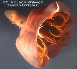
Title Illustrations

| Scientific Name | Stauroteuthis syrtensis |
|---|---|
| Location | 2560 ft depth in Oceanographer canyon, off Gulf of Maine USA |
| Comments | Collected by the manned submersible "Johnson-Sea-Link-II and photographed in a shipboard aquarium. |
| Specimen Condition | Live Specimen |
| View | Side |
| Copyright | © 2005 Per R. Flood |
About This Page
Martin Collins

Aberdeen University, Aberdeen, UK
University of Hawaii, Honolulu, HI, USA
National Museum of Natural History, Washington, D. C. , USA
Page copyright © 2016 and
 Page: Tree of Life
Stauroteuthidae . Stauroteuthis .
Authored by
Martin Collins, Richard E. Young, and Michael Vecchione.
The TEXT of this page is licensed under the
Creative Commons Attribution-NonCommercial License - Version 3.0. Note that images and other media
featured on this page are each governed by their own license, and they may or may not be available
for reuse. Click on an image or a media link to access the media data window, which provides the
relevant licensing information. For the general terms and conditions of ToL material reuse and
redistribution, please see the Tree of Life Copyright
Policies.
Page: Tree of Life
Stauroteuthidae . Stauroteuthis .
Authored by
Martin Collins, Richard E. Young, and Michael Vecchione.
The TEXT of this page is licensed under the
Creative Commons Attribution-NonCommercial License - Version 3.0. Note that images and other media
featured on this page are each governed by their own license, and they may or may not be available
for reuse. Click on an image or a media link to access the media data window, which provides the
relevant licensing information. For the general terms and conditions of ToL material reuse and
redistribution, please see the Tree of Life Copyright
Policies.
- Content changed 28 April 2008
Citing this page:
Collins, Martin, Richard E. Young, and Michael Vecchione. 2008. Stauroteuthidae . Stauroteuthis . Version 28 April 2008 (under construction). http://tolweb.org/Stauroteuthis/20092/2008.04.28 in The Tree of Life Web Project, http://tolweb.org/


