Carinariidae
Roger R. Seapy


This tree diagram shows the relationships between several groups of organisms.
The root of the current tree connects the organisms featured in this tree to their containing group and the rest of the Tree of Life. The basal branching point in the tree represents the ancestor of the other groups in the tree. This ancestor diversified over time into several descendent subgroups, which are represented as internal nodes and terminal taxa to the right.

You can click on the root to travel down the Tree of Life all the way to the root of all Life, and you can click on the names of descendent subgroups to travel up the Tree of Life all the way to individual species.
For more information on ToL tree formatting, please see Interpreting the Tree or Classification. To learn more about phylogenetic trees, please visit our Phylogenetic Biology pages.
close boxIntroduction
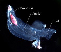
The bodies of carinariids, like those of pterotracheids, are greatly enlarged by comparison with the microscopic atlantids. Also like the pterotracheids, the body in most species is elongated and cylindrical, and is divisible into three regions (proboscis, trunk and tail). A reduced shell is present, growing out of the larval shell after metamorphosis. The adult shell is either cap-shaped, covering the viscera, gonads, heart and gills (Carinaria and Pterosoma) or is microscopic, modified only slightly from the larval shell, and embedded dorsally in the visceral nucleus (Cardiapoda). A fin sucker is present in both sexes (with the exception of Cardiapoda richardi, which lacks a sucker in females) and the size of the sucker relative to the swimming fin is very small in comparison with the atlantids.
Brief Diagnosis
Heteropod molluscs with:
- Body elongated and basically cylindrical, divided into proboscis, trunk and tail
- Viscera compacted into a stalked visceral nucleus
- Shell cap-shaped, covering the viscera and other organs, or microscopic, embedded in the dorsal part of the visceral nucleus.
Characteristics
- Body morphology
- Body elongate and basically cylindrical, except in Pterosoma which has a broad and disc-shaped trunk
- Like pterotracheids, body divided into proboscis, trunk and tail regions
- Visceral nucleus dorsal and stalked
- Shell
- Adult shell macroscopic in Carinaria (cap-shaped) and Pterosoma (flattened) and covering the viscera and gills, or microscopic in Cardiapoda and embedded in dorsal tissues of the visceral nucleus
 Click on an image to view larger version & data in a new window
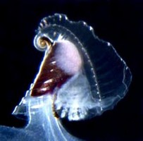
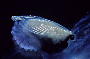
Figure. Stalked visceral nucleus and shell in Carinaria galea (left) and Pterosoma planum (right). In the left image, the digestive gland is dark brown and testis is pink. ©
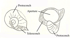
Figure. Drawings of the microscopic adult shell in Cardiapoda placenta, viewed from the right side and aperture. Shell diameter (left image) about 0.8 mm. Drawings modified from Tesch (1949).
- Protoconch located at apex of adult shell (teleoconch) in Carinaria and Pterosoma
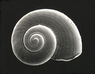
- Larval shell globular; umbilicus open with radiating striae; aperture oval with smooth outer lip
 Click on an image to view larger version & data in a new window
 Click on an image to view larger version & data in a new window
Figure. Larval shell of Carinaria galea. Shell viewed from right side, left side, and aperture, respectively. Note the striae radiating from the umbilicus (middle image) and the operculum inside the shell aperture (right image). Scale bar = 250 µm (applies to all images). ©
- Adult shell macroscopic in Carinaria (cap-shaped) and Pterosoma (flattened) and covering the viscera and gills, or microscopic in Cardiapoda and embedded in dorsal tissues of the visceral nucleus
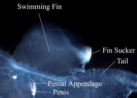
- Swimming fin and sucker
- Fin sucker small and located on the posteroventral margin of the swimming fin; present in both sexes
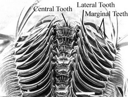
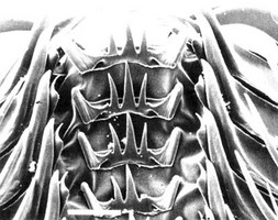
- Radula
- Shape broadly triangular, with a relative low and limited number of tooth rows (34-44)
- Central (rachidian) tooth broad with a low base, three cusps, and prominent posterolateral processes
- Lateral teeth with a small, curved cusp on the inner accessory plate
- Marginal teeth monocuspid
Comments
Most species are in the genus Carinaria; Pterosoma is monotypic and there are two species of Cardiapoda. The genera can be distinguished by the following characters:
| Genus | Shell size and shape | Shell location |
|---|---|---|
| Carinaria | Macroscopic; cap-shaped, laterally compressed | Covers the tall, stalked visceral nucleus |
| Pterosoma | Macroscopic; flattened, oblong in dorsal view | Covers the low, stalked visceral nucleus |
| Cardiapoda | Microscopic; adult shell shield-shaped extension of the larval shell | Imbedded in dorsal tissues of the visceral nucleus |
References
Lalli, C. M. and R. W. Gilmer. 1989. Pelagic snails. The biology of holoplanktonic gastropod snails. Stanford University Press, Stanford. 259 pp.
Richter, G. and R. R. Seapy. 1999. Heteropoda, pp. 621-647. In: D. Boltovskoy (ed.), South Atlantic Zooplankton. Backhuys Publishers, Leiden.
Seapy, R. R. and C. Thiriot-Quiévreux. 1994. Veliger larvae of Carinariidae (Mollusca: Heteropoda) from Hawaiian waters. Veliger 37: 336-343.
Tesch, J. J. 1949. Heteropoda. Dana Report 34, 53 pp., 5 plates.
Title Illustrations

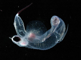
| Scientific Name | Carinaria japonica |
|---|---|
| Location | Hawaiian waters |
| Specimen Condition | Live Specimen |
| Sex | Male |
| Life Cycle Stage | adult |
| View | right side |
| Image Use |
 This media file is licensed under the Creative Commons Attribution-NonCommercial License - Version 3.0. This media file is licensed under the Creative Commons Attribution-NonCommercial License - Version 3.0.
|
| Copyright |
©

|
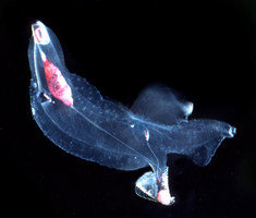
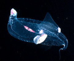
| Scientific Name | Cardiapoda richardi |
|---|---|
| Location | Sargasso Sea |
| Specimen Condition | Live Specimen |
| Sex | Female |
| Life Cycle Stage | adult |
| View | ventral |
| Copyright | © L. Madin |
| Scientific Name | Pterosoma planum |
|---|---|
| Location | Hawaiian waters |
| Specimen Condition | Live Specimen |
| Life Cycle Stage | adult |
| View | ventro-lateral |
| Image Use |
 This media file is licensed under the Creative Commons Attribution-NonCommercial License - Version 3.0. This media file is licensed under the Creative Commons Attribution-NonCommercial License - Version 3.0.
|
| Copyright |
©

|
About This Page

California State University, Fullerton, California, USA
Correspondence regarding this page should be directed to Roger R. Seapy at
Page copyright © 2007
 Page: Tree of Life
Carinariidae .
Authored by
Roger R. Seapy.
The TEXT of this page is licensed under the
Creative Commons Attribution License - Version 3.0. Note that images and other media
featured on this page are each governed by their own license, and they may or may not be available
for reuse. Click on an image or a media link to access the media data window, which provides the
relevant licensing information. For the general terms and conditions of ToL material reuse and
redistribution, please see the Tree of Life Copyright
Policies.
- First online 13 July 2007
- Content changed 05 December 2007
Citing this page:
Seapy, Roger R. 2007. Carinariidae . Version 05 December 2007 (under construction). http://tolweb.org/Carinariidae/28733/2007.12.05 in The Tree of Life Web Project, http://tolweb.org/





 Go to quick links
Go to quick search
Go to navigation for this section of the ToL site
Go to detailed links for the ToL site