Atlanta tokiokai
Roger R. SeapyIntroduction
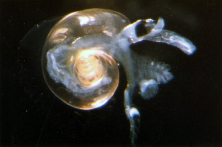
Atlanta tokiokai is a small species (to 2.5-3.0 mm). The shell is light yellowish-brown, and the inside of the outer lip is brown. The keel is tall and rounded in side profile. The spire has a globose shape with an apical angle of about 80°. The spire consists of about 5-1/2 whorls, with sutures that are very shallow and difficult to distinguish when the spire is viewed from the side. The spire bears prominent tubercles that are arranged in distinct spiral lines; in some cases they can be sufficiently elevated to give the appearance of spiral ridges. The inner wall of the spire (viewed with transmitted light) has radially-arranged lines that can be difficult to resolve because of the strongly developed surface sculpture. The spire whorls are decalcified internally, as in A. inclinata. On the left side of the shell the whorl adjacent to the umbilicus bears a prominent ridge, and between the ridge and the umbilicus the whorl is flattened. Eyes type b, operculum type c, and radula type II, with the number of tooth rows limited to about 60. The radula is small and ribbon-like, with a growth angle of about 9°, and the lateral teeth have an accessory cusp. The species has a cosmopolitan distribution in tropical to subtropical waters.Diagnosis
- Shell small; maximal diameter = 2.5-3.0 mm
- Shell light yellowish-brown; inside of outer lip brown
- Keel tall and rounded
- Spire shape globose, with an apical angle of about 80°
- Spire of about 5-1/2 whorls, with very shallow sutures
- Spire whorls decalcified internally
- Spire with prominent tubercles arranged in spiral rows
- Inner surface of spire with radially-arranged lines
- Whorl adjacent to umbilicus with prominent ridge; whorl surface flattened between ridge and umbilicus
- Eyes type b
- Operculum type c
- Radula type II
- Number of tooth rows in radula limited to about 60
- Radula small and ribbon-like, with growth angle of about 9°
- Lateral teeth bear an accessory cusp
Characteristics
- Shell
- Shell small, with a maximal diameter of 2.5 to 3.0 mm (Tesch, 1906 [identified as A. inclinata]); to 3.0 mm (Richter, 1990); and, to 2.6 mm (Seapy, 1990)
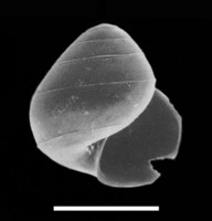
- Shell coloration light yellowish-brown (see title illustration)
- Keel tall and rounded in side profile
 Click on an image to view larger version & data in a new window
 Click on an image to view larger version & data in a new window
 Click on an image to view larger version & data in a new window
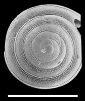
Figure. Shell of Atlanta tokiokai, with views of the right side (left) and the spire (right). Images from Richter (1990, figs. 1 and 13), modified by addition of scale bars (= 1.0 mm and 100 µm, respectively). © 1990 G. Richter
- Spire globose in shape, with an apical angle of about 80° (see SEM below)
- Spire consists of about 5-1/2 whorls
- Spire sutures very shallow, with the result that when the spire is viewed from the side (as in the larval shell above) adjacent whorls are difficult to distinguish from each other
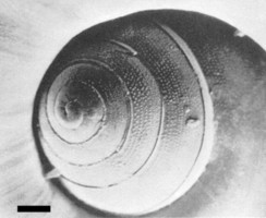
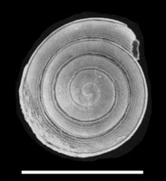
- Spire surface with well-developed tubercles (or punctae) that are arranged in spiral rows (click on first SEM below for detail); in some cases the tubercles can be raised and appear to form spiral ridges (click on second SEM below for detail)
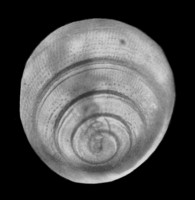
- Inner walls of spire whorls with radially-arranged lines, which can only be resolved using transmitted light (see SEM below). Note that the prominent tubercles on the spire whorls somewhat obscure the underlying lines
- Internal spire walls decalcified (viewed using transmitted light, see the SEM below)
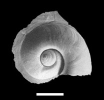
- Whorl adjacent to the umbilicus with a prominent ridge. The portion of the whorl medial to the ridge is flattened (see second SEM below)
 Click on an image to view larger version & data in a new window
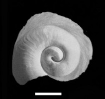
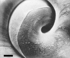
Figure. Left side of shell of Atlanta tokiokai, with views of entire shell (left) and the umbilical region (right). Images from Richter (1990, figs. 2 and 16), modified by addition of scale bars (= 1.0 and 100 µm, respectively). © 1990 G. Richter
- Shell small, with a maximal diameter of 2.5 to 3.0 mm (Tesch, 1906 [identified as A. inclinata]); to 3.0 mm (Richter, 1990); and, to 2.6 mm (Seapy, 1990)
- Eyes type b
- Operculum type c
- Radula type II
- Number of tooth rows comprising radula about 60
- Radula small with a ribbon-like shape; growth angle about 9°
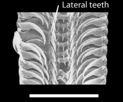
- Lateral teeth with an accessory cusp (see SEM below; click on image to see detail)
 Click on an image to view larger version & data in a new window
Figure. Section of radula from Atlanta tokiokai, viewed dorsally (from lower portion of radula photographed above). Image from Richter (1990, fig. 31), modified by addition of label and scale bar (= 100 µm). © 1990 G. Richter
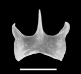
- Rachidian teeth (from adult portion of radula) about 20% narrower than in A. inclinata. In turn, the rachidian teeth in A. tokiokai are about 40% narrower than in either A. gibbosa or A. meteori (Richter, 1990)
- Number of tooth rows comprising radula about 60
Comments
Atlanta tokiokai was described by van der Spoel and Troost in 1972. Their description was based on a single specimen collected from the Caribbean Sea, North Atlantic Ocean. The shell appears to be an early post-metamorphic one; note in the second drawing the upturning of the last whorl behind the aperture that indicates the redirection of the axis of shell growth seen in the adult shell.


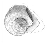
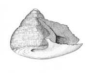
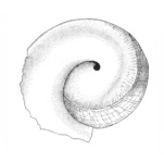

Figure. Drawings of the holotype shell of Atlanta tokiokai, from the right side (left), in apertural view (middle), and umbilical region on the left side (right). Shell diameter = 2.3 mm. © 1972 van der Spoel & Troost
References
Richter, G. 1990. Zur Kenntnis der Gattung Atlanta (IV). Die Atlanta inclinata-Gruppe (Prosobranchia: Heteropoda). Archiv für Molluskenkunde 119: 239-275.
Richter, G. and R. R. Seapy. 1999. Heteropoda, pp. 621-647. In: D. Boltovskoy (ed.), South Atlantic Zooplankton. Backhuys Publishers, Leiden.
Seapy, R. R. 1990. The pelagic family Atlantidae (Gastropoda, Heteropoda) from Hawaiian waters: a faunistic survey. Malacologia 32: 107-130.
Tesch, J. J. 1906. Die Heteropoden der Siboga-Expedition. Monograph 51, 112 pp, 14 plates. E. J. Brill, Leiden.
Spoel, S. van der and D. G. Troost. 1972. Atlanta tokiokai, a new heteropod (Gastropoda). Basteria 36: 1-6.
About This Page

California State University, Fullerton, California, USA
Correspondence regarding this page should be directed to Roger R. Seapy at
Page copyright © 2011
 Page: Tree of Life
Atlanta tokiokai .
Authored by
Roger R. Seapy.
The TEXT of this page is licensed under the
Creative Commons Attribution License - Version 3.0. Note that images and other media
featured on this page are each governed by their own license, and they may or may not be available
for reuse. Click on an image or a media link to access the media data window, which provides the
relevant licensing information. For the general terms and conditions of ToL material reuse and
redistribution, please see the Tree of Life Copyright
Policies.
- First online 01 July 2010
- Content changed 23 July 2011
Citing this page:
Seapy, Roger R. 2011. Atlanta tokiokai . Version 23 July 2011 (under construction). http://tolweb.org/Atlanta_tokiokai/28772/2011.07.23 in The Tree of Life Web Project, http://tolweb.org/






 Go to quick links
Go to quick search
Go to navigation for this section of the ToL site
Go to detailed links for the ToL site